Horii Toshihiro, Fujioka Tomoyuki, Takahashi Marie, Mori Mio, Tsuchiya Junichi, Yamaga Emi, Yamada Hirofumi, Kimura Mizuki, Kishino Mitsuhiro, Tateishi Ukihide
Department of Diagnostic Radiology, Tokyo Medical and Dental University, 1-5-45 Yushima, Bunkyo-ku, 113 - 8510, Tokyo, Japan.
Radiol Case Rep. 2020 Dec;15(12):2560-2564. doi: 10.1016/j.radcr.2020.09.036. Epub 2020 Sep 23.
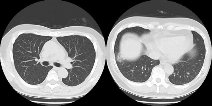
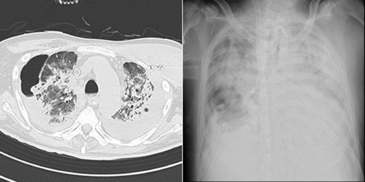
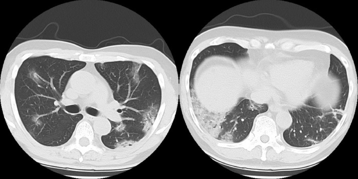
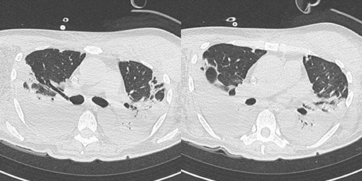
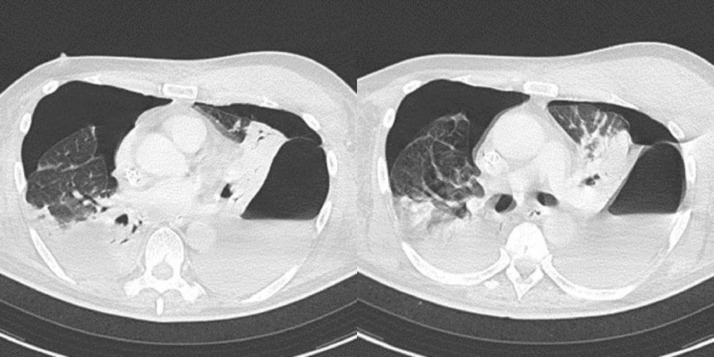
Coronavirus disease 2019 (COVID-19) has become a major threat to public health since the outbreak in Wuhan in 2019. Chest computed tomography is recommended for COVID-19 cases for evaluation and follow up of pneumonia and related complication. We report the case of a 66-year-old man with underlying hypertension and a history of smoking 76 packs a year; he was frequently monitored by computed tomography for pulmonary changes during the period from early symptom onset to death. Furthermore, he developed a pneumothorax during the course. The occurrence of pneumothorax in COVID-19 patients is not common, and there have been only a few previous reports. This is a valuable case of pneumothorax in a patient with COVID-19 treated with a ventilator and extracorporeal membrane oxygenation. This case and previous reports suggest that pneumothorax occurs in COVID-19 with a relatively late onset (3-8 weeks). Long-term pneumonia morbidity, steroid therapy, positive pressure ventilation, and extracorporeal membrane oxygenation can cause pneumothorax, leading to capillary and alveolar damage.
自2019年在武汉爆发以来,2019冠状病毒病(COVID-19)已成为对公众健康的重大威胁。对于COVID-19病例,建议进行胸部计算机断层扫描,以评估和随访肺炎及相关并发症。我们报告了一例66岁男性病例,该患者患有高血压,有每年吸烟76包的吸烟史;从症状初发到死亡期间,他经常接受计算机断层扫描以监测肺部变化。此外,他在病程中发生了气胸。COVID-19患者发生气胸并不常见,之前仅有少数报道。这是一例使用呼吸机和体外膜肺氧合治疗的COVID-19患者发生气胸的有价值病例。该病例及之前的报道表明,COVID-19患者气胸发病相对较晚(3 - 8周)。长期肺炎发病率、类固醇治疗、正压通气和体外膜肺氧合可导致气胸,进而导致毛细血管和肺泡损伤。