Jacobs Michael A, Umbricht Christopher B, Parekh Vishwa S, El Khouli Riham H, Cope Leslie, Macura Katarzyna J, Harvey Susan, Wolff Antonio C
The Russell H. Morgan Department of Radiology and Radiological Science, The Johns Hopkins School of Medicine, Baltimore, MD 21205, USA.
Sidney Kimmel Comprehensive Cancer Center, The Johns Hopkins School of Medicine, Baltimore, MD 21205, USA.
Cancers (Basel). 2020 Sep 27;12(10):2772. doi: 10.3390/cancers12102772.
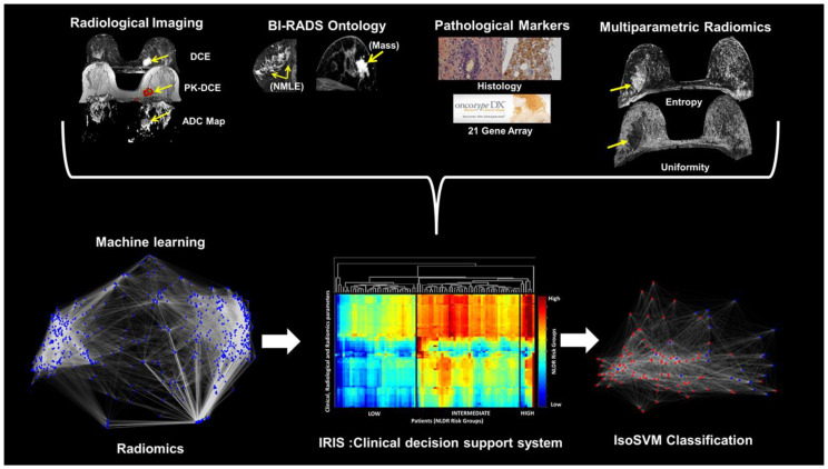
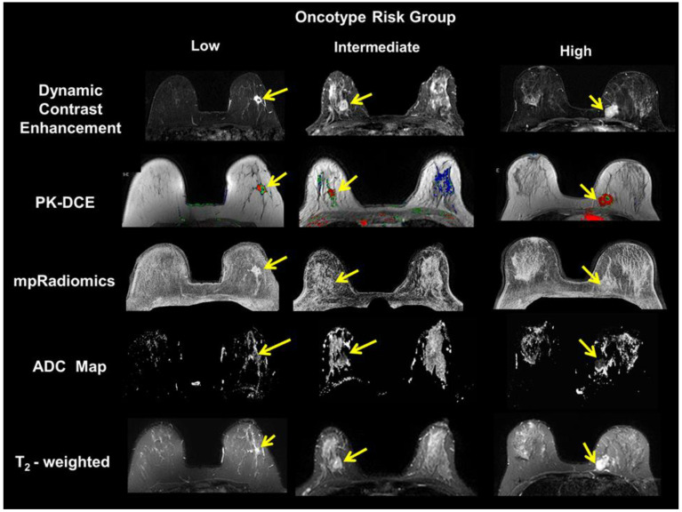
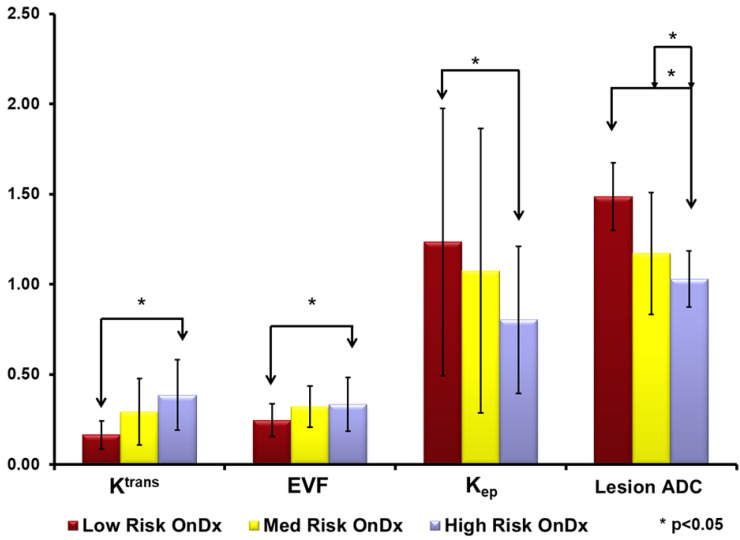
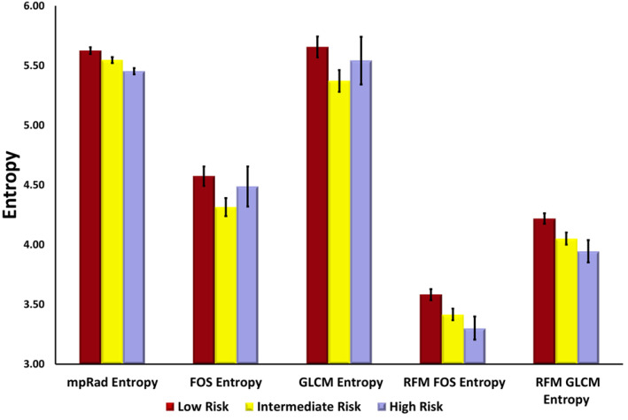
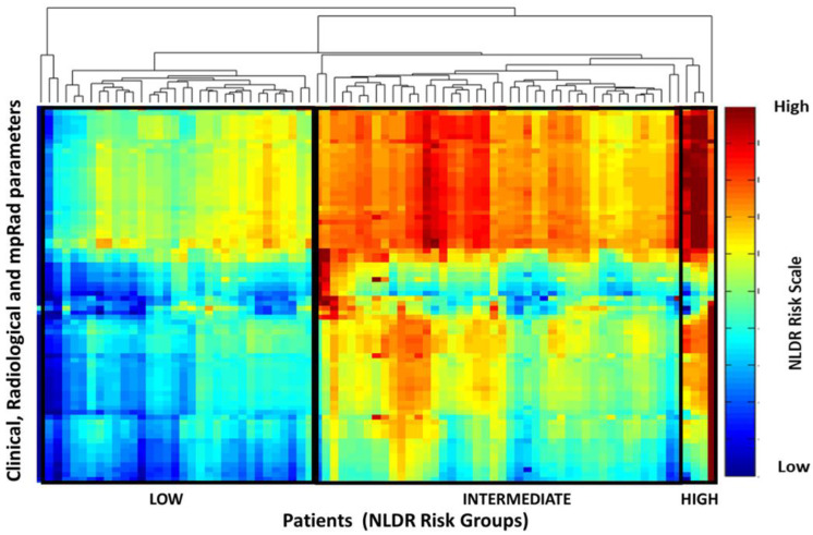
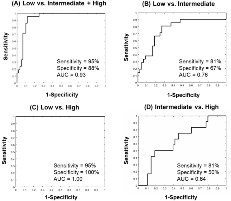
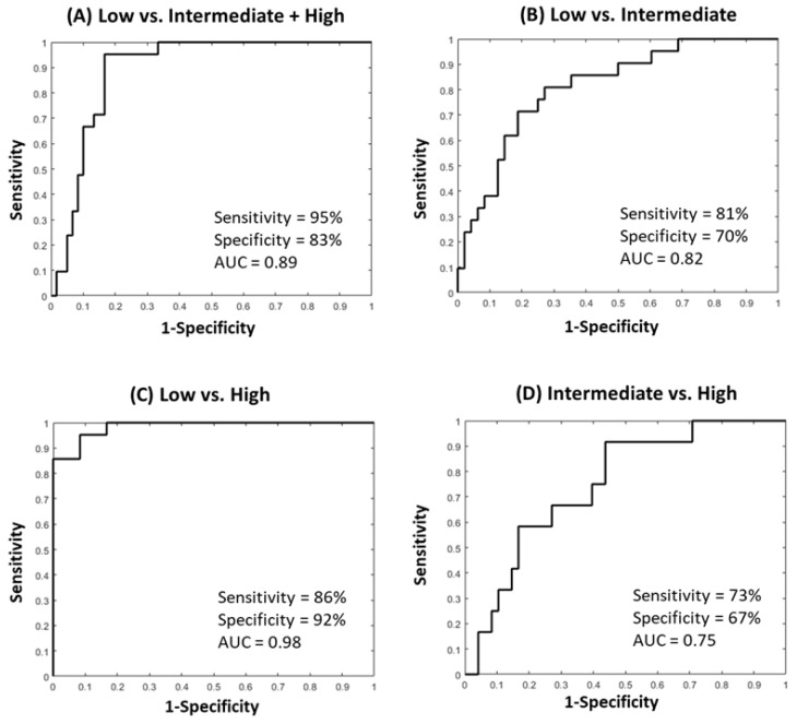
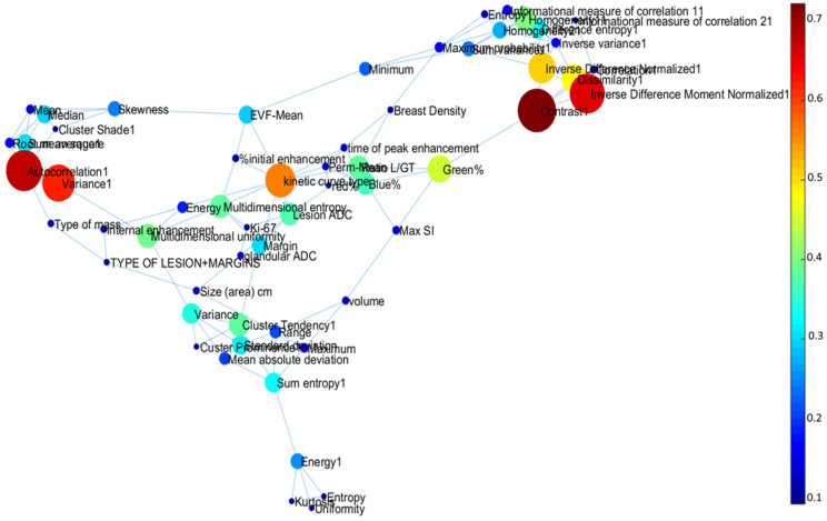
Optimal use of multiparametric magnetic resonance imaging (mpMRI) can identify key MRI parameters and provide unique tissue signatures defining phenotypes of breast cancer. We have developed and implemented a new machine-learning informatic system, termed Informatics Radiomics Integration System (IRIS) that integrates clinical variables, derived from imaging and electronic medical health records (EHR) with multiparametric radiomics (mpRad) for identifying potential risk of local or systemic recurrence in breast cancer patients. We tested the model in patients ( = 80) who had Estrogen Receptor positive disease and underwent OncotypeDX gene testing, radiomic analysis, and breast mpMRI. The IRIS method was trained using the mpMRI, clinical, pathologic, and radiomic descriptors for prediction of the OncotypeDX risk score. The trained mpRad IRIS model had a 95% and specificity was 83% with an Area Under the Curve (AUC) of 0.89 for classifying low risk patients from the intermediate and high-risk groups. The lesion size was larger for the high-risk group (2.9 ± 1.7 mm) and lower for both low risk (1.9 ± 1.3 mm) and intermediate risk (1.7 ± 1.4 mm) groups. The lesion apparent diffusion coefficient (ADC) map values for high- and intermediate-risk groups were significantly ( < 0.05) lower than the low-risk group (1.14 vs. 1.49 × 10 mm/s). These initial studies provide deeper insight into the clinical, pathological, quantitative imaging, and radiomic features, and provide the foundation to relate these features to the assessment of treatment response for improved personalized medicine.
多参数磁共振成像(mpMRI)的优化使用能够识别关键的MRI参数,并提供定义乳腺癌表型的独特组织特征。我们开发并实施了一种新的机器学习信息系统,称为信息放射组学整合系统(IRIS),该系统将源自影像学和电子医疗健康记录(EHR)的临床变量与多参数放射组学(mpRad)相结合,以识别乳腺癌患者局部或全身复发的潜在风险。我们在80例雌激素受体阳性疾病且接受了OncotypeDX基因检测、放射组学分析和乳腺mpMRI的患者中测试了该模型。IRIS方法使用mpMRI、临床、病理和放射组学描述符进行训练,以预测OncotypeDX风险评分。经过训练的mpRad IRIS模型在将低风险患者与中高风险组进行分类时,灵敏度为95%,特异性为83%,曲线下面积(AUC)为0.89。高风险组的病变大小更大(2.9±1.7毫米),低风险组(1.9±1.3毫米)和中风险组(1.7±1.4毫米)的病变大小较小。高风险组和中风险组的病变表观扩散系数(ADC)图值显著低于低风险组(<0.05)(1.14对1.49×10毫米²/秒)。这些初步研究为临床、病理、定量成像和放射组学特征提供了更深入的见解,并为将这些特征与治疗反应评估相关联以改善个性化医疗奠定了基础。