The Russell H. Morgan Department of Radiology and Radiological Science, The Johns Hopkins School of Medicine, Baltimore, MD, 21205, USA.
Department of Computer Science, The Johns Hopkins University, Baltimore, MD, 21208, USA.
Breast Cancer Res Treat. 2020 Apr;180(2):407-421. doi: 10.1007/s10549-020-05533-5. Epub 2020 Feb 4.
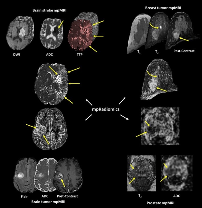
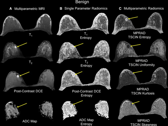
Multiparametric radiological imaging is vital for detection, characterization, and diagnosis of many different diseases. Radiomics provide quantitative metrics from radiological imaging that may infer potential biological meaning of the underlying tissue. However, current methods are limited to regions of interest extracted from a single imaging parameter or modality, which limits the amount of information available within the data. This limitation can directly affect the integration and applicable scope of radiomics into different clinical settings, since single image radiomics are not capable of capturing the true underlying tissue characteristics in the multiparametric radiological imaging space. To that end, we developed a multiparametric imaging radiomic (mpRad) framework for extraction of first and second order radiomic features from multiparametric radiological datasets.
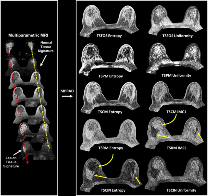
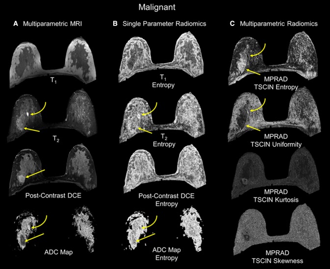
We developed five different radiomic techniques that extract different aspects of the inter-voxel and inter-parametric relationships within the high-dimensional multiparametric magnetic resonance imaging breast datasets. Our patient cohort consisted of 138 breast patients, where, 97 patients had malignant lesions and 41 patients had benign lesions. Sensitivity, specificity, receiver operating characteristic (ROC) and areas under the curve (AUC) analysis were performed to assess diagnostic performance of the mpRad parameters. Statistical significance was set at p < 0.05.
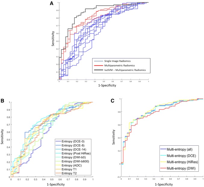
The mpRad features successfully classified malignant from benign breast lesions with excellent sensitivity and specificity of 82.5% and 80.5%, respectively, with Area Under the receiver operating characteristic Curve (AUC) of 0.87 (0.81-0.93). mpRad provided a 9-28% increase in AUC metrics over single radiomic parameters.
We have introduced the mpRad framework that extends radiomic analysis from single images to multiparametric datasets for better characterization of the underlying tissue biology.
多参数放射影像学对于多种不同疾病的检测、特征描述和诊断至关重要。放射组学可从放射影像学中提取定量指标,这些指标可能推断出潜在的组织生物学意义。然而,目前的方法仅限于从单个成像参数或模态提取感兴趣区域,这限制了数据中可用信息的数量。这种局限性直接影响了放射组学在不同临床环境中的整合和适用范围,因为单图像放射组学无法捕捉多参数放射影像学空间中真实的潜在组织特征。为此,我们开发了一种多参数成像放射组学(mpRad)框架,用于从多参数放射影像学数据集中提取一阶和二阶放射组学特征。
我们开发了五种不同的放射组学技术,从高维多参数磁共振成像乳腺数据集中提取体素间和参数间关系的不同方面。我们的患者队列包括 138 名乳腺患者,其中 97 名患者患有恶性病变,41 名患者患有良性病变。进行了敏感性、特异性、接受者操作特征(ROC)和曲线下面积(AUC)分析,以评估 mpRad 参数的诊断性能。统计显著性设置为 p<0.05。
mpRad 特征成功地将恶性与良性乳腺病变进行分类,敏感性和特异性分别为 82.5%和 80.5%,ROC 曲线下面积(AUC)为 0.87(0.81-0.93)。mpRad 提供了比单放射组学参数高 9-28%的 AUC 指标。
我们引入了 mpRad 框架,将放射组学分析从单图像扩展到多参数数据集,以更好地描述潜在的组织生物学。