Human Health Science Graduate School of Medicine Kyoto University Kyoto Japan.
Department of Systems Science Graduate School of Informatics Kyoto University Kyoto Japan.
J Am Heart Assoc. 2020 Oct 20;9(19):e016422. doi: 10.1161/JAHA.120.016422. Epub 2020 Sep 30.
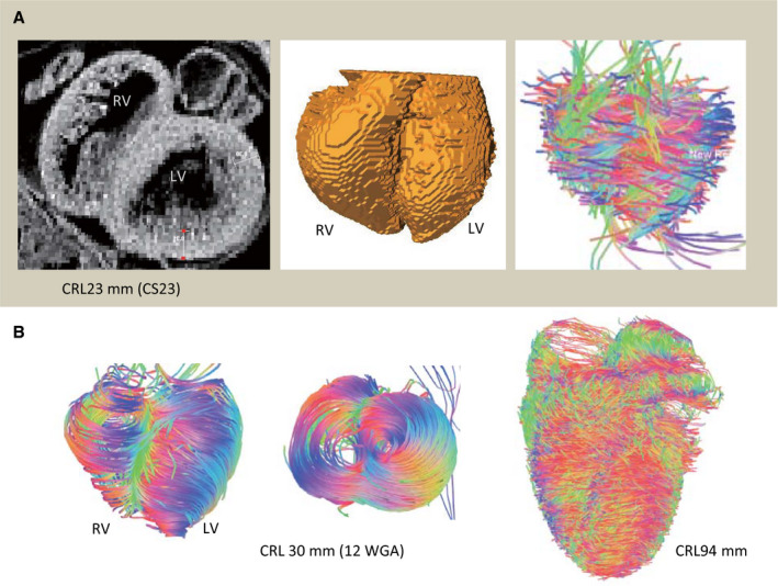
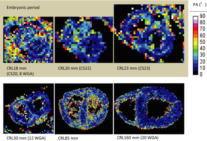
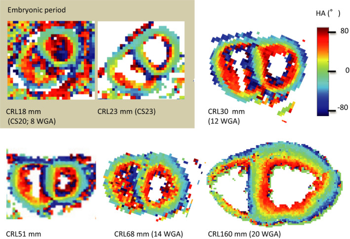
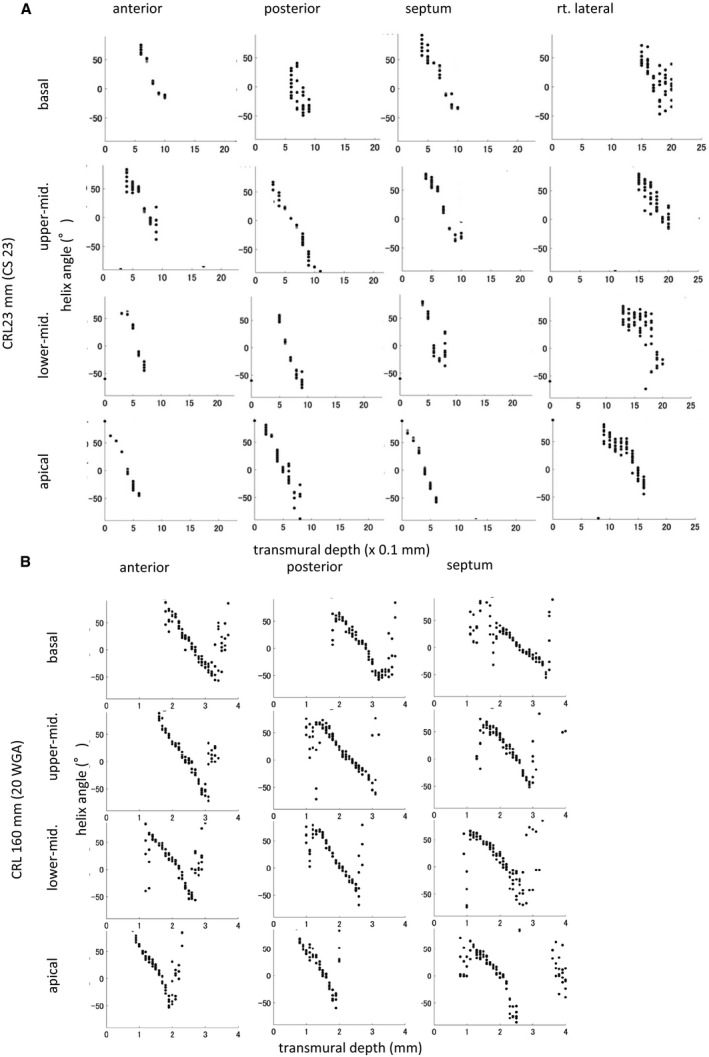
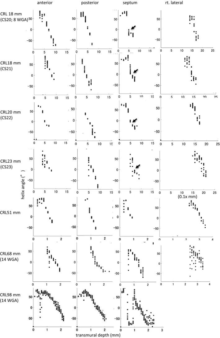
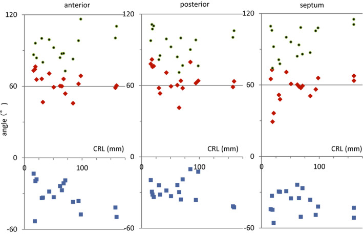
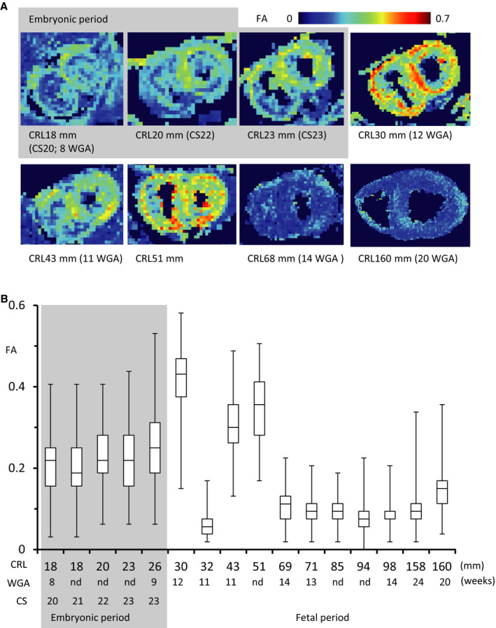
Background Detection of the fiber orientation pattern of the myocardium using diffusion tensor magnetic resonance imaging lags ≈12 weeks of gestational age (WGA) behind fetal myocardial remodeling with invasion by the developing coronary vasculature (8 WGA). We aimed to use diffusion tensor magnetic resonance imaging tractography to characterize the evolution of fiber architecture in the developing human heart from the later embryonic period. Methods and Results Twenty human specimens (8-24 WGA) from the Kyoto Collection of Human Embryos and Fetuses, including specimens from the embryonic period (Carnegie stages 20-23), were used. Diffusion tensor magnetic resonance imaging data were acquired with a 7T magnetic resonance system. Fractional anisotropy and helix angle were calculated using standard definitions. In all samples, the fibers ran helically in an organized pattern in both the left and right ventricles. A smooth transmural change in helix angle values (from positive to negative) was detected in all 16 directions of the ventricles. This feature was observed in almost all small (Carnegie stage 23) and large samples. A higher fractional anisotropy value was detected at the outer side of the anterior wall and septum at Carnegie stage 20 to 22, which spread around the ventricular wall at Carnegie stage 23 and in the early fetal samples (11-12 WGA). The fractional anisotropy value of the left ventricular walls decreased in samples with ≥13 WGA, which remained low (≈0.09) in larger samples. Conclusions From the human late embryonic period (from 8 WGA), the helix angle arrangement of the myocardium is comparable to that of the adult, indicating that the myocardial structure blueprint, organization, and integrity are already formed.
背景 心肌纤维方向模式的弥散张量磁共振成像检测落后于胎儿心肌重塑(胚胎第 8 周),而心肌重塑是由正在发育的冠状动脉侵入引起的。我们旨在使用弥散张量磁共振成像示踪术来描述从胚胎后期到人类心脏纤维结构的演变。
方法和结果 从京都人类胚胎和胎儿收集组织中使用 20 个人类标本(8-24 周龄),包括胚胎期(卡内基阶段 20-23)的标本。使用 7T 磁共振系统采集弥散张量磁共振成像数据。使用标准定义计算各向异性分数和螺旋角。在所有样本中,纤维在左心室和右心室中均呈螺旋状排列。在心室的 16 个方向上,均检测到螺旋角值(从正到负)的平滑壁间变化。这一特征在几乎所有的小(卡内基阶段 23)和大样本中都能观察到。在卡内基阶段 20 到 22 期间,在前壁和间隔的外侧检测到较高的各向异性分数值,在卡内基阶段 23 及在早期胎儿样本(11-12 周龄)中,该值在心室壁周围扩散。左心室壁的各向异性分数值在≥13 周龄的样本中降低,在较大的样本中仍保持较低(≈0.09)。
结论 从人类胚胎后期(8 周龄)开始,心肌的螺旋角排列与成人相似,表明心肌结构蓝图、组织和完整性已经形成。