Schnorrenberg Sebastian, Ghareeb Hassan, Frahm Lars, Grotjohann Tim, Jensen Nickels, Teichmann Thomas, Hell Stefan W, Lipka Volker, Jakobs Stefan
Department of NanoBiophotonics Max Planck Institute for Biophysical Chemistry Göttingen Germany.
Department of Plant Cell Biology Albrecht-von-Haller Institute of Plant Sciences University of Göttingen Göttingen Germany.
Plant Direct. 2020 Sep 3;4(9):e00261. doi: 10.1002/pld3.261. eCollection 2020 Sep.
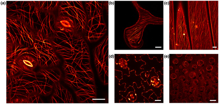
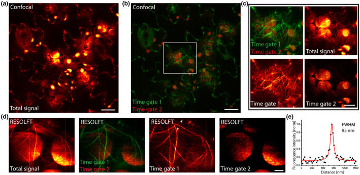
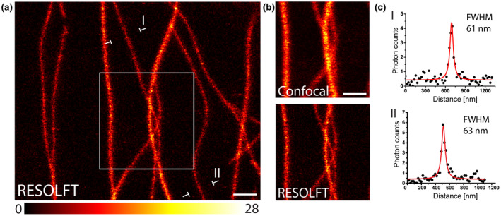
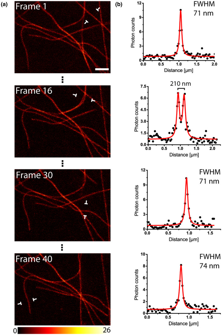
Subdiffraction super-resolution fluorescence microscopy, or nanoscopy, has seen remarkable developments in the last two decades. Yet, for the visualization of plant cells, nanoscopy is still rarely used. In this study, we established RESOLFT nanoscopy on living green plant tissue. Live-cell RESOLFT nanoscopy requires and utilizes comparatively low light doses and intensities to overcome the diffraction barrier. We generated a transgenic plant line expressing the reversibly switchable fluorescent protein rsEGFP2 fused to the mammalian microtubule-associated protein 4 (MAP4) in order to ubiquitously label the microtubule cytoskeleton. We demonstrate the use of RESOLFT nanoscopy for extended time-lapse imaging of cortical microtubules in leaf discs. By combining our approach with fluorescence lifetime gating, we were able to acquire live-cell RESOLFT images even close to chloroplasts, which exhibit very strong autofluorescence. The data demonstrate the feasibility of subdiffraction resolution imaging in transgenic plant material with minimal requirements for sample preparation.
亚衍射超分辨率荧光显微镜,即纳米显微镜,在过去二十年中取得了显著进展。然而,对于植物细胞的可视化,纳米显微镜仍很少被使用。在本研究中,我们在绿色植物活体组织上建立了RESOLFT纳米显微镜技术。活细胞RESOLFT纳米显微镜需要并利用相对较低的光剂量和强度来克服衍射障碍。我们构建了一个转基因植物品系,该品系表达与哺乳动物微管相关蛋白4(MAP4)融合的可逆开关荧光蛋白rsEGFP2,以便对微管细胞骨架进行普遍标记。我们展示了RESOLFT纳米显微镜在叶片圆盘皮层微管长时间延时成像中的应用。通过将我们的方法与荧光寿命门控相结合,我们甚至能够在靠近叶绿体的区域获取活细胞RESOLFT图像,叶绿体具有非常强的自发荧光。数据证明了在转基因植物材料中进行亚衍射分辨率成像的可行性,且对样品制备的要求极低。