Lab. of Bioassays and Cellular Dynamics, Department of Chemical and Biological Sciences, Institute of Biosciences, UNESP-São Paulo State University, 18618-970, Botucatu, São Paulo, Brazil.
School of Dentistry, University of Taubaté, 12020-340, Taubaté, São Paulo, Brazil.
Biomed Res Int. 2020 Sep 13;2020:3026893. doi: 10.1155/2020/3026893. eCollection 2020.
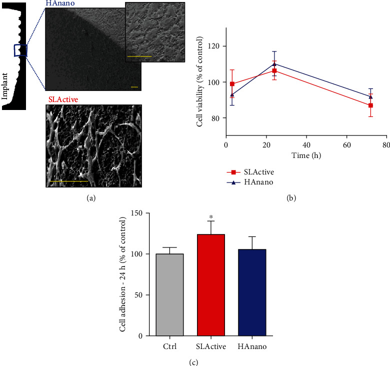
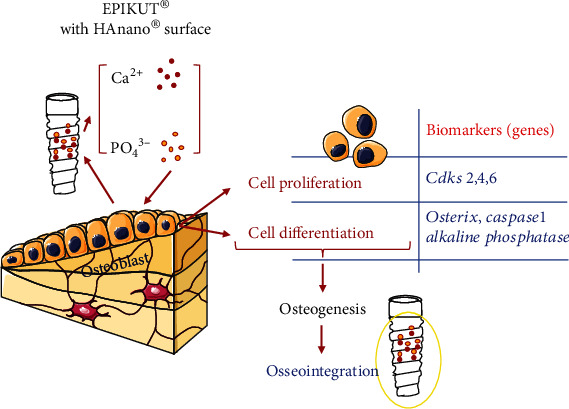
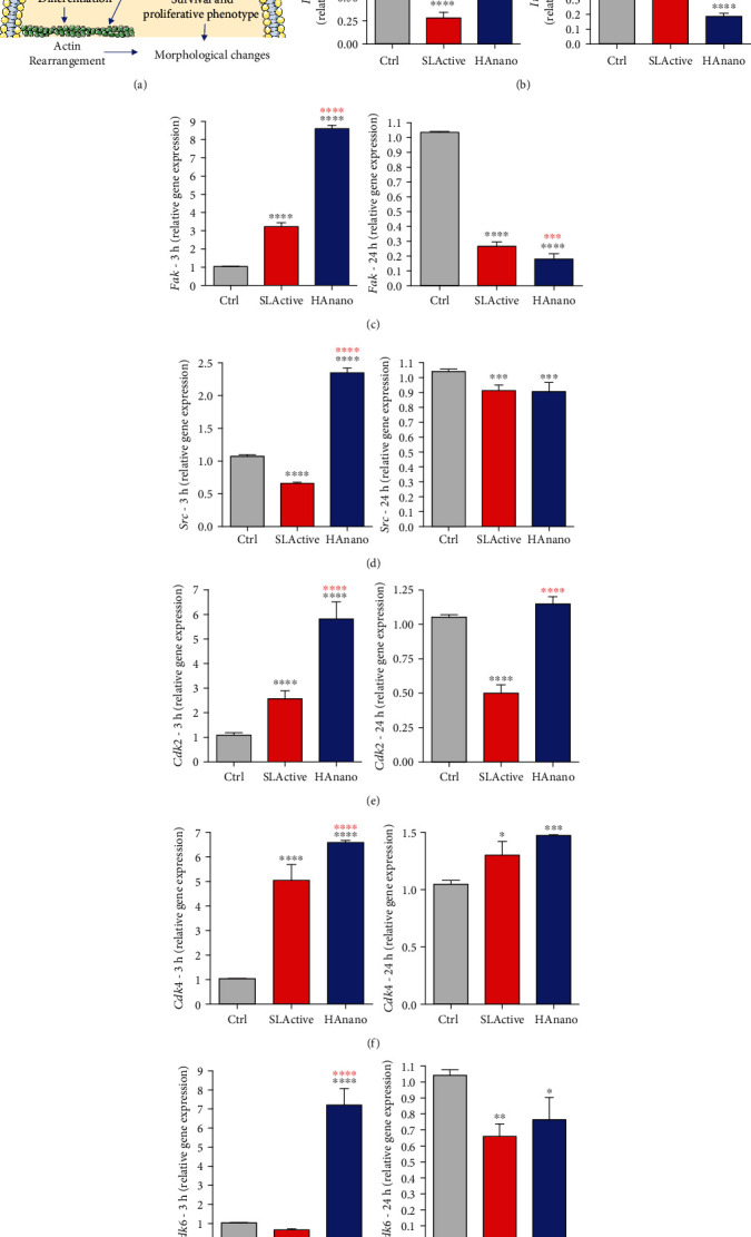
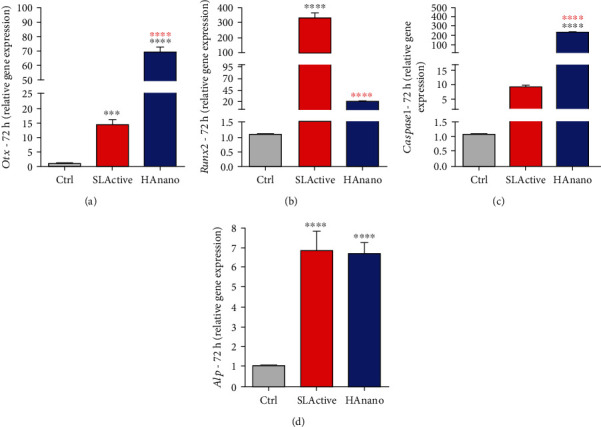
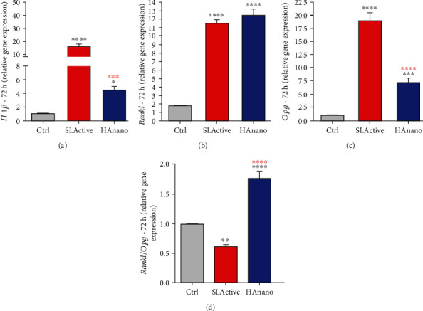
There is an increased effort on developing novel and active surfaces in order to accelerate their osteointegration, such as nanosized crystalline hydroxyapatite coating (HAnano®). To better understand the biological behavior of osteoblasts grown on HAnano® surface, the set of data was compared with SLActive®, a hydrophilic sandblasted titanium surface. Methodologically, osteoblasts were seeded on both surfaces up to 72 hours, to allow evaluating cell adhesion, viability, and set of genes encoding proteins related with adhesion, proliferation, and differentiation. Our data shows HAnano® displays an interesting substrate to support cell adhesion with typical spread morphologic cells, while SLActive®-adhering cells presented fusiform morphology. Our data shows that the cellular adhesion mechanism was accompanied with upexpression of , , and , favoring the assembling of focal adhesion platforms and coupling cell cycle progression (upmodulating of , , and genes) in response to HAnano®. Additionally, both bioactive surfaces promoted osteoblast differentiation stimulus, by activating , , and genes. Although both surfaces promoted gene expression, Opg gene expression was higher in SLActive® and this difference reflected on the / ratio. Finally, Caspase1 gene was significantly upmodulated in response to HAnano® and it suggests an involvement of the inflammasome complex. Collectively, this study provides enough evidences to support that the nanohydroxyapatite-coated surface provides the necessary microenvironment to drive osteoblast performance on dental implants and these stages of osteogenesis are expected during the early stages of osseointegration.
为了加速其骨整合,人们正在努力开发新型活性表面,例如纳米级结晶羟基磷灰石涂层(HAnano®)。为了更好地了解在 HAnano®表面上生长的成骨细胞的生物学行为,将该数据集与 SLActive®(亲水喷砂钛表面)进行了比较。从方法学上讲,将成骨细胞接种到两种表面上,直到 72 小时,以评估细胞黏附,活力和编码与黏附,增殖和分化相关的蛋白质的一组基因。我们的数据表明,HAnano®显示出一种有趣的基质,可以支持细胞黏附,形成典型的展开形态细胞,而 SLActive®附着的细胞则呈现出梭形形态。我们的数据表明,细胞黏附机制伴随着上调基因的表达,从而有利于形成粘着斑平台,并在响应 HAnano®时使细胞周期进程偶联(上调基因的表达)。此外,两种生物活性表面都通过激活基因的表达来促进成骨细胞分化刺激。尽管两种表面都促进了基因的表达,但在 SLActive®中 Opg 基因的表达更高,这种差异反映在/比率上。最后,Caspase1 基因的表达在响应 HAnano®时显著上调,这表明炎症小体复合物的参与。总的来说,这项研究提供了足够的证据支持纳米羟基磷灰石涂层表面提供了驱动牙种植体上成骨细胞性能的必要微环境,并且这些成骨阶段预计将在骨整合的早期阶段发生。