Won Jungeun, Huang Pin-Chieh, Boppart Stephen A
Department of Bioengineering, University of Illinois at Urbana-Champaign, Urbana, IL.
Beckman Institute for Advanced Science and Technology, University of Illinois at Urbana-Champaign, Urbana, IL.
JPhys Photonics. 2020 Jul;2(3). doi: 10.1088/2515-7647/ab8a59. Epub 2020 May 15.
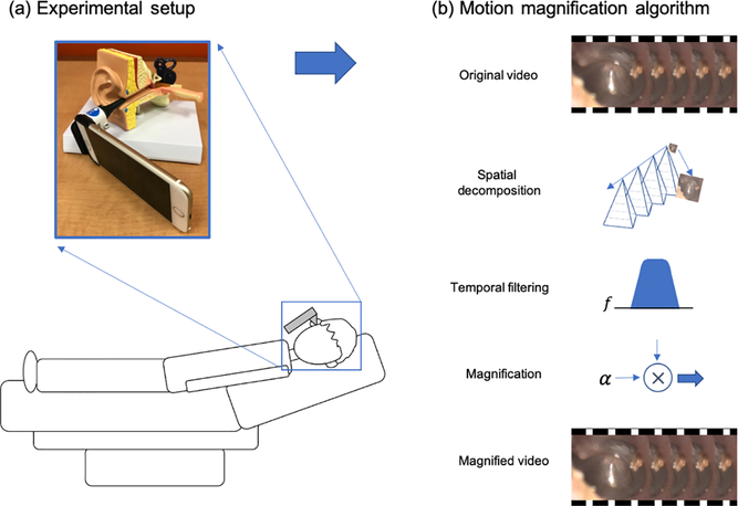
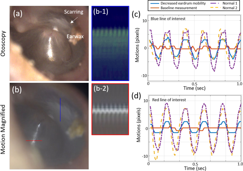
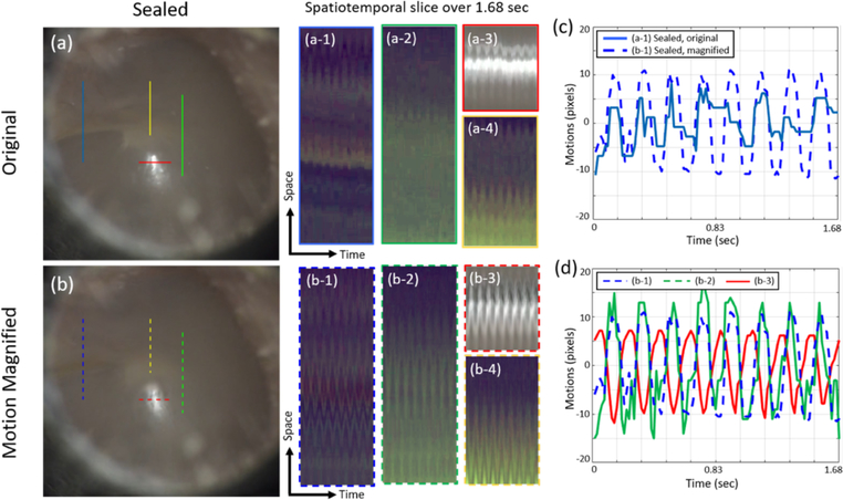
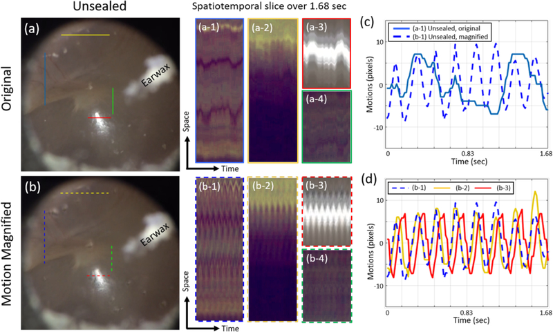
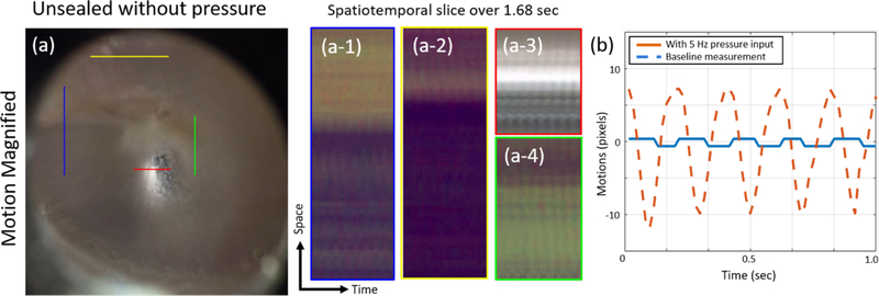
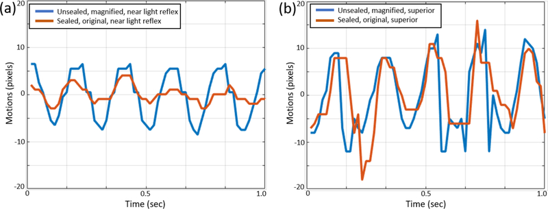
Pneumatic otoscopy is the recommended diagnostic method for middle ear infections. Physicians use a pneumatic otoscope to assess the position of the eardrum (bulging or retraction) as well as the eardrum mobility while an insufflation bulb is squeezed to generate air pressure changes in a sealed ear canal. While pneumatic otoscopy provides increased sensitivity and specificity by detecting decreased eardrum mobility, there exist many challenges to correctly perform and interpret results. For example, the ear canal must be sealed using a specialized ear speculum to deliver sufficiently large pressure changes that can induce visible movements of an eardrum. To overcome this challenge, video motion magnification is proposed to amplify pneumatic-induced motions of the eardrum without sealing of the ear canal. Pneumatic otoscopy is performed on adult subjects using a smartphone camera with an otoscope attachment at 60 frames per second, with pressure inputs at 5 Hz. Phase-based Eulerian motion magnification is applied to magnify spatiotemporal dependent motions in the video. As a result, the motion magnification of unsealed pneumatic otoscopy reveals comparable eardrum motions as in standard pneumatic otoscopy with a sealed ear canal. Furthermore, the estimated motions (in pixels) are quantified to examine the spatial and the temporal variations of the eardrum motions. The motion magnification may avoid the need for sealing the ear canal as well as decrease patient discomfort in pneumatic otoscopy, improving the capability and the usability as a point-of-care diagnostic tool in primary care and otology.
鼓气耳镜检查是诊断中耳感染的推荐方法。医生使用鼓气耳镜评估鼓膜的位置(膨出或内陷)以及鼓膜的活动度,同时挤压打气球以在密封的耳道内产生气压变化。虽然鼓气耳镜检查通过检测鼓膜活动度降低提高了敏感性和特异性,但正确执行和解读结果存在许多挑战。例如,必须使用专门的耳镜来密封耳道,以产生足够大的压力变化,从而引起鼓膜的可见运动。为了克服这一挑战,有人提出使用视频运动放大技术来放大鼓膜的鼓气诱导运动,而无需密封耳道。使用带有耳镜附件的智能手机摄像头以每秒60帧的速度对成年受试者进行鼓气耳镜检查,压力输入频率为5赫兹。应用基于相位的欧拉运动放大技术来放大视频中时空相关的运动。结果,未密封鼓气耳镜检查的运动放大显示出与密封耳道的标准鼓气耳镜检查中相当的鼓膜运动。此外,对估计的运动(以像素为单位)进行量化,以检查鼓膜运动的空间和时间变化。运动放大技术可以避免在鼓气耳镜检查中密封耳道的需要,并减少患者的不适,提高其作为初级保健和耳科学即时诊断工具的能力和可用性。