Farzaneh Negar, Williamson Craig A, Jiang Cheng, Srinivasan Ashok, Bapuraj Jayapalli R, Gryak Jonathan, Najarian Kayvan, Soroushmehr S M Reza
Department of Computational Medicine and Bioinformatics, University of Michigan, Ann Arbor, MI 48109, USA.
Michigan Center for Integrative Research in Critical Care (MCIRCC), University of Michigan, Ann Arbor, MI 48109, USA.
Diagnostics (Basel). 2020 Sep 30;10(10):773. doi: 10.3390/diagnostics10100773.
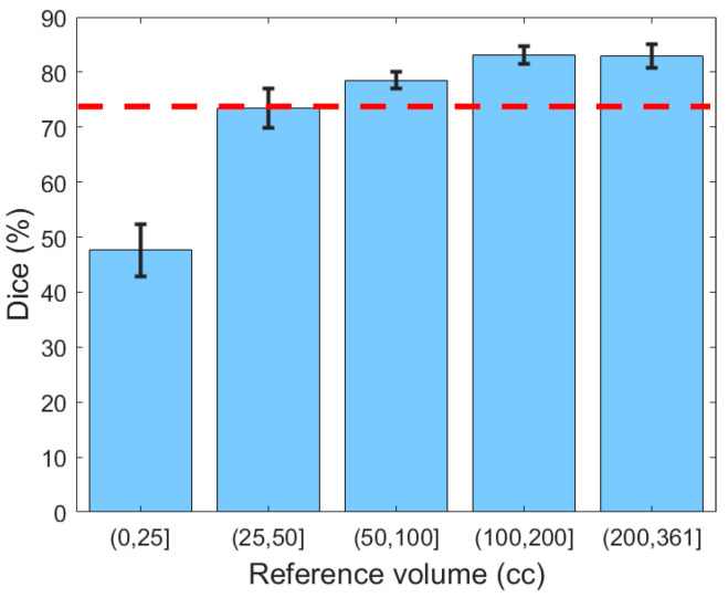
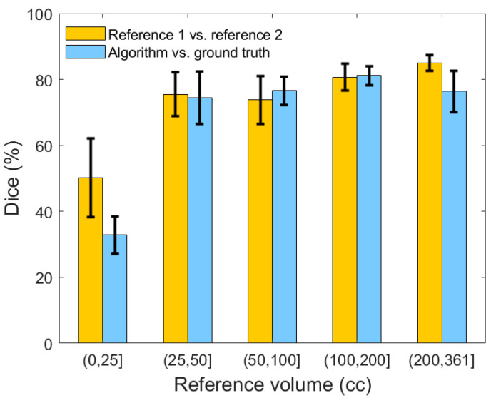
Detection and severity assessment of subdural hematoma is a major step in the evaluation of traumatic brain injuries. This is a retrospective study of 110 computed tomography (CT) scans from patients admitted to the Michigan Medicine Neurological Intensive Care Unit or Emergency Department. A machine learning pipeline was developed to segment and assess the severity of subdural hematoma. First, the probability of each point belonging to the hematoma region was determined using a combination of hand-crafted and deep features. This probability provided the initial state of the segmentation. Next, a 3D post-processing model was applied to evolve the initial state and delineate the hematoma. The recall, precision, and Dice similarity coefficient of the proposed segmentation method were 78.61%, 76.12%, and 75.35%, respectively, for the entire population. The Dice similarity coefficient was 79.97% for clinically significant hematomas, which compared favorably to an inter-rater Dice similarity coefficient. In volume-based severity analysis, the proposed model yielded an F1, recall, and specificity of 98.22%, 98.81%, and 92.31%, respectively, in detecting moderate and severe subdural hematomas based on hematoma volume. These results show that the combination of classical image processing and deep learning can outperform deep learning only methods to achieve greater average performance and robustness. Such a system can aid critical care physicians in reducing time to intervention and thereby improve long-term patient outcomes.
硬膜下血肿的检测与严重程度评估是创伤性脑损伤评估的重要步骤。这是一项对密歇根大学医学神经重症监护病房或急诊科收治患者的110份计算机断层扫描(CT)进行的回顾性研究。开发了一种机器学习流程来分割硬膜下血肿并评估其严重程度。首先,使用手工特征和深度特征的组合来确定每个点属于血肿区域的概率。该概率提供了分割的初始状态。接下来,应用三维后处理模型来演化初始状态并勾勒出血肿。对于整个人群,所提出的分割方法的召回率、精确率和Dice相似系数分别为78.61%、76.12%和75.35%。对于具有临床意义的血肿,Dice相似系数为79.97%,与评分者间的Dice相似系数相比具有优势。在基于体积的严重程度分析中,所提出的模型在根据血肿体积检测中度和重度硬膜下血肿时,F1值、召回率和特异性分别为98.22%、98.81%和92.31%。这些结果表明,经典图像处理与深度学习相结合的方法比仅使用深度学习的方法表现更优,能够实现更高的平均性能和更强的鲁棒性。这样的系统可以帮助重症监护医生减少干预时间,从而改善患者的长期预后。