• 文献检索
  • 文档翻译
  • 深度研究
  • 学术资讯
  • Suppr Zotero 插件Zotero 插件
  • 邀请有礼
  • 套餐&价格
  • 历史记录
应用&插件
Suppr Zotero 插件Zotero 插件浏览器插件Mac 客户端Windows 客户端微信小程序
定价
高级版会员购买积分包购买API积分包
服务
文献检索文档翻译深度研究API 文档MCP 服务
关于我们
关于 Suppr公司介绍联系我们用户协议隐私条款
关注我们

Suppr 超能文献

核心技术专利:CN118964589B侵权必究
粤ICP备2023148730 号-1Suppr @ 2026

文献检索

告别复杂PubMed语法,用中文像聊天一样搜索,搜遍4000万医学文献。AI智能推荐,让科研检索更轻松。

立即免费搜索

文件翻译

保留排版,准确专业,支持PDF/Word/PPT等文件格式,支持 12+语言互译。

免费翻译文档

深度研究

AI帮你快速写综述,25分钟生成高质量综述,智能提取关键信息,辅助科研写作。

立即免费体验

肺部超声作为 SARS-CoV-2 感染的诊断工具。

Lung ultrasound as diagnostic tool for SARS-CoV-2 infection.

机构信息

Department of Emergency Medicine, COVID Care Unit, Santa Maria Delle Grazie Hospital, Via Domitiana, 5, 80078, Pozzuoli, Naples, Italy.

Department of Translational Medical Sciences, Federico II University, Naples, Italy.

出版信息

Intern Emerg Med. 2021 Mar;16(2):471-476. doi: 10.1007/s11739-020-02512-y. Epub 2020 Oct 3.

DOI:10.1007/s11739-020-02512-y
PMID:33011929
原文链接:https://pmc.ncbi.nlm.nih.gov/articles/PMC7532928/
Abstract

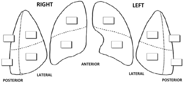
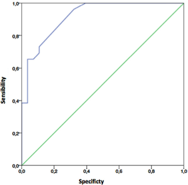
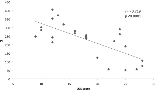
The aim of this study was to explore the role of lung ultrasound (LUS) in the diagnosis of SARS-CoV-2 infection and to verify its utility in the prediction of lung disease's severity and outcome. Fifty-three consecutive patients presenting to the Emergency Department of Santa Maria delle Grazie Hospital with high suspicion of SARS-CoV-2 infection underwent diagnostic test for SARS-CoV-2 on samples obtained from nasopharyngeal swab as well as complete proper diagnostic work-up that included clinical evaluation, laboratory tests, blood gas analyses, chest CT and LUS. A semiquantitative analysis of B-lines distribution was performed to calculate the LUS score. Patients were divided into two groups according to the results of both SARS-CoV-2 diagnostic test and other exams (Group A = pneumonia due to SARS-CoV2 infection vs Group B = no SARS-CoV2 infection and another definite diagnosis). LUS showed an excellent accuracy in predicting the diagnosis of SARS-CoV-2 infection (area under the ROC curve of 0.92 with a sensibility of 73% and a specificity of 89% a the cut-off of 12.5). LUS score was more impaired in SARS-CoV-2 patients (18.1 ± 6.0 vs 7.6 ± 5.9, p < 0.00001) and it is significantly negatively correlated with PF ratio values (r = - 0.719, p < 0.0001). An intrahospital mortality rate of 46% was found; patients with adverse outcome had significant higher value of LUS, PF, LDH, and APACHE II score. None of these parameters was predictive of mortality. LUS is a useful tool for the early detection of SARS-CoV-2 infection and for the evaluation of the disease severity, but does not predict mortality. Further studies with repeated evaluations of LUS score are needed to further explore the role of LUS in the assessment of severity in SARS-CoV-2 disease and in the monitoring of the response to treatments.

摘要

本研究旨在探讨肺部超声(LUS)在 SARS-CoV-2 感染诊断中的作用,并验证其在预测肺部疾病严重程度和结局方面的效用。53 例连续就诊于圣玛丽亚德拉格拉齐亚医院急诊科,高度怀疑 SARS-CoV-2 感染的患者,对鼻咽拭子样本进行 SARS-CoV-2 诊断检测,并进行完整的适当诊断检查,包括临床评估、实验室检查、血气分析、胸部 CT 和 LUS。对 B 线分布进行半定量分析,以计算 LUS 评分。根据 SARS-CoV-2 诊断检测和其他检查的结果,将患者分为两组(A 组=SARS-CoV-2 感染引起的肺炎与 B 组=无 SARS-CoV-2 感染和其他明确诊断)。LUS 在预测 SARS-CoV-2 感染的诊断方面具有出色的准确性(ROC 曲线下面积为 0.92,敏感度为 73%,特异性为 89%,截断值为 12.5)。SARS-CoV-2 患者的 LUS 评分更差(18.1±6.0 与 7.6±5.9,p<0.00001),与 PF 比值呈显著负相关(r=-0.719,p<0.0001)。院内死亡率为 46%;不良结局患者的 LUS、PF、LDH 和 APACHE II 评分显著较高。这些参数均不能预测死亡率。LUS 是早期发现 SARS-CoV-2 感染和评估疾病严重程度的有用工具,但不能预测死亡率。需要进一步研究重复评估 LUS 评分,以进一步探讨 LUS 在评估 SARS-CoV-2 疾病严重程度和监测治疗反应中的作用。

https://cdn.ncbi.nlm.nih.gov/pmc/blobs/d4bd/7532928/44190b5e9fde/11739_2020_2512_Fig3_HTML.jpg
https://cdn.ncbi.nlm.nih.gov/pmc/blobs/d4bd/7532928/8f692d8b1c99/11739_2020_2512_Fig1_HTML.jpg
https://cdn.ncbi.nlm.nih.gov/pmc/blobs/d4bd/7532928/651d6d4aa72c/11739_2020_2512_Fig2_HTML.jpg
https://cdn.ncbi.nlm.nih.gov/pmc/blobs/d4bd/7532928/44190b5e9fde/11739_2020_2512_Fig3_HTML.jpg
https://cdn.ncbi.nlm.nih.gov/pmc/blobs/d4bd/7532928/8f692d8b1c99/11739_2020_2512_Fig1_HTML.jpg
https://cdn.ncbi.nlm.nih.gov/pmc/blobs/d4bd/7532928/651d6d4aa72c/11739_2020_2512_Fig2_HTML.jpg
https://cdn.ncbi.nlm.nih.gov/pmc/blobs/d4bd/7532928/44190b5e9fde/11739_2020_2512_Fig3_HTML.jpg

相似文献

1
Lung ultrasound as diagnostic tool for SARS-CoV-2 infection.肺部超声作为 SARS-CoV-2 感染的诊断工具。
Intern Emerg Med. 2021 Mar;16(2):471-476. doi: 10.1007/s11739-020-02512-y. Epub 2020 Oct 3.
2
Comparative study of lung ultrasound and chest computed tomography scan in the assessment of severity of confirmed COVID-19 pneumonia.肺超声与胸部 CT 扫描在评估确诊 COVID-19 肺炎严重程度中的对比研究。
Intensive Care Med. 2020 Sep;46(9):1707-1713. doi: 10.1007/s00134-020-06186-0. Epub 2020 Jul 29.
3
Lung Ultrasound in the Emergency Department for Early Identification of COVID-19 Pneumonia.急诊科肺部超声在早期识别 COVID-19 肺炎中的应用。
Respiration. 2021;100(2):145-153. doi: 10.1159/000512782. Epub 2020 Dec 7.
4
Correlation between Chest Computed Tomography and Lung Ultrasonography in Patients with Coronavirus Disease 2019 (COVID-19).新型冠状病毒肺炎(COVID-19)患者的胸部计算机断层扫描与肺部超声的相关性。
Ultrasound Med Biol. 2020 Nov;46(11):2918-2926. doi: 10.1016/j.ultrasmedbio.2020.07.003. Epub 2020 Jul 13.
5
Lung Ultrasound in COVID-19 Pneumonia: Correlations with Chest CT on Hospital admission.COVID-19 肺炎的肺部超声:与入院时胸部 CT 的相关性。
Respiration. 2020;99(7):617-624. doi: 10.1159/000509223. Epub 2020 Jun 22.
6
Lung ultrasound in the emergency department - a valuable tool in the management of patients presenting with respiratory symptoms during the SARS-CoV-2 pandemic.急诊科肺部超声 - SARS-CoV-2 大流行期间,处理呼吸系统症状患者的有价值工具。
BMC Emerg Med. 2020 Dec 7;20(1):96. doi: 10.1186/s12873-020-00389-w.
7
Prognostic value of bedside lung ultrasound score in patients with COVID-19.COVID-19 患者床边肺部超声评分的预后价值。
Crit Care. 2020 Dec 22;24(1):700. doi: 10.1186/s13054-020-03416-1.
8
The role of lung ultrasound as a frontline diagnostic tool in the era of COVID-19 outbreak.肺部超声在 COVID-19 疫情时代作为一线诊断工具的作用。
Intern Emerg Med. 2021 Apr;16(3):749-756. doi: 10.1007/s11739-020-02524-8. Epub 2020 Oct 22.
9
Low Sensitivity of Admission Lung US Compared to Chest CT for Diagnosis of Lung Involvement in a Cohort of 82 Patients with COVID-19 Pneumonia.在82例新冠肺炎患者队列中,与胸部CT相比,入院时肺部超声对诊断肺部受累的敏感性较低。
Medicina (Kaunas). 2021 Mar 4;57(3):236. doi: 10.3390/medicina57030236.
10
Lung Ultrasound May Support Diagnosis and Monitoring of COVID-19 Pneumonia.肺部超声检查可能有助于 COVID-19 肺炎的诊断和监测。
Ultrasound Med Biol. 2020 Nov;46(11):2908-2917. doi: 10.1016/j.ultrasmedbio.2020.07.018. Epub 2020 Jul 20.

引用本文的文献

1
Pulmonary ultrasound in COVID-19 and non-COVID-19 pneumonia in South Africa: An observational study.南非新冠肺炎与非新冠肺炎肺炎的肺部超声:一项观察性研究。
Afr J Thorac Crit Care Med. 2025 Mar 28;31(1):e1887. doi: 10.7196/AJTCCM.2025.v31i1.1887. eCollection 2025.
2
Lung Ultrasound Score in COVID-19 Patients Correlates with PO/FiO, Intubation Rates, and Mortality.新冠肺炎患者的肺部超声评分与 PO/FiO、插管率和死亡率相关。
West J Emerg Med. 2024 Jan;25(1):28-39. doi: 10.5811/westjem.59975.
3
Italian Society of Anesthesia, Analgesia, Resuscitation, and Intensive Care expert consensus statement on the use of lung ultrasound in critically ill patients with coronavirus disease 2019 (ITACO).

本文引用的文献

1
Non-invasive CPAP in mild and moderate ARDS secondary to SARS-CoV-2.COVID-19 相关轻度和中度 ARDS 患者的无创 CPAP 治疗。
Respir Physiol Neurobiol. 2020 Sep;280:103489. doi: 10.1016/j.resp.2020.103489. Epub 2020 Jul 3.
2
Eculizumab treatment in patients with COVID-19: preliminary results from real life ASL Napoli 2 Nord experience.COVID-19 患者的依库珠单抗治疗:来自 ASL Napoli 2 Nord 真实经验的初步结果。
Eur Rev Med Pharmacol Sci. 2020 Apr;24(7):4040-4047. doi: 10.26355/eurrev_202004_20875.
3
Routine blood tests as a potential diagnostic tool for COVID-19.
意大利麻醉、镇痛、复苏与重症监护学会关于2019冠状病毒病(COVID-19)重症患者肺部超声应用的专家共识声明(ITACO)。
J Anesth Analg Crit Care. 2021 Nov 24;1(1):16. doi: 10.1186/s44158-021-00015-6.
4
Lung ultrasound score severity cut-off points in COVID-19 pneumonia. A systematic review and validating cohort.新型冠状病毒肺炎中肺部超声评分的严重程度截断点:一项系统评价及验证队列研究
Med Clin (Engl Ed). 2023 Jun 23;160(12):531-539. doi: 10.1016/j.medcle.2023.01.023. Epub 2023 Jun 16.
5
Lung ultrasound score severity cut-off points in COVID-19 pneumonia. A systematic review and validating cohort.COVID-19 肺炎肺超声评分严重程度截断值:系统评价和验证队列研究。
Med Clin (Barc). 2023 Jun 23;160(12):531-539. doi: 10.1016/j.medcli.2023.01.024. Epub 2023 Mar 10.
6
Utility and diagnostic test properties of pulmonary and cardiovascular point of care ultra-sonography (POCUS) in COVID-19 patients admitted to critical care unit.重症监护病房收治的COVID-19患者中肺部和心血管床旁超声检查(POCUS)的效用及诊断测试特性
Eur J Radiol Open. 2022;9:100451. doi: 10.1016/j.ejro.2022.100451. Epub 2022 Nov 3.
7
Lung ultrasound national report (LUNARE PROJECT) by the Italian Society of Internal Medicine (SIMI).意大利内科医学会(SIMI)发布的肺部超声全国报告(LUNARE项目)。
Intern Emerg Med. 2023 Mar;18(2):685-688. doi: 10.1007/s11739-022-03149-9. Epub 2022 Nov 3.
8
Point-of-care Lung Ultrasound, Lung CT and NEWS to Predict Adverse Outcomes and Mortality in COVID-19 Associated Pneumonia.床旁肺部超声、肺部 CT 和 NEWS 预测 COVID-19 相关肺炎的不良结局和死亡率。
J Intensive Care Med. 2022 Dec;37(12):1614-1624. doi: 10.1177/08850666221111731. Epub 2022 Nov 1.
9
Reliability and clinical correlations of semi-quantitative lung ultrasound on BLUE points in COVID-19 mechanically ventilated patients: The 'BLUE-LUSS'-A feasibility clinical study.COVID-19 机械通气患者 BLUE 点半定量肺部超声的可靠性及临床相关性:“BLUE-LUSS”-A 可行性临床研究。
PLoS One. 2022 Oct 14;17(10):e0276213. doi: 10.1371/journal.pone.0276213. eCollection 2022.
10
[Korean Clinical Imaging Guidelines for Justification of Diagnostic Imaging Study for COVID-19].[韩国关于新型冠状病毒肺炎诊断影像检查合理性的临床影像指南]
Taehan Yongsang Uihakhoe Chi. 2022 Mar;83(2):265-283. doi: 10.3348/jksr.2021.0117. Epub 2022 Jan 6.
常规血液检查可作为 COVID-19 的潜在诊断工具。
Clin Chem Lab Med. 2020 Jun 25;58(7):1095-1099. doi: 10.1515/cclm-2020-0398.
4
Thoracic ultrasound and SARS-COVID-19: a pictorial essay.胸部超声与新型冠状病毒肺炎:图文并茂的文章
J Ultrasound. 2020 Jun;23(2):217-221. doi: 10.1007/s40477-020-00458-7. Epub 2020 Apr 16.
5
The epidemiology and clinical information about COVID-19.新型冠状病毒肺炎的流行病学和临床信息。
Eur J Clin Microbiol Infect Dis. 2020 Jun;39(6):1011-1019. doi: 10.1007/s10096-020-03874-z. Epub 2020 Apr 14.
6
COVID-19 pneumonia: different respiratory treatments for different phenotypes?新冠肺炎:针对不同表型采用不同的呼吸治疗方法?
Intensive Care Med. 2020 Jun;46(6):1099-1102. doi: 10.1007/s00134-020-06033-2. Epub 2020 Apr 14.
7
COVID-19 pneumonia: A review of typical CT findings and differential diagnosis.COVID-19 肺炎:CT 表现的典型特征及鉴别诊断综述。
Diagn Interv Imaging. 2020 May;101(5):263-268. doi: 10.1016/j.diii.2020.03.014. Epub 2020 Apr 3.
8
COVID-19 pneumonia manifestations at the admission on chest ultrasound, radiographs, and CT: single-center study and comprehensive radiologic literature review.COVID-19肺炎入院时胸部超声、X光片及CT表现:单中心研究及放射学文献综述
Eur J Radiol Open. 2020;7:100231. doi: 10.1016/j.ejro.2020.100231. Epub 2020 Apr 4.
9
Our Italian experience using lung ultrasound for identification, grading and serial follow-up of severity of lung involvement for management of patients with COVID-19.我们在意大利利用肺部超声对新冠肺炎患者进行肺部受累情况的识别、分级及严重程度的连续随访以进行管理的经验。
Echocardiography. 2020 Apr;37(4):625-627. doi: 10.1111/echo.14664. Epub 2020 Apr 15.
10
The Clinical and Chest CT Features Associated With Severe and Critical COVID-19 Pneumonia.与严重和危重新冠肺炎相关的临床和胸部 CT 特征。
Invest Radiol. 2020 Jun;55(6):327-331. doi: 10.1097/RLI.0000000000000672.