Zhou Shi-Chong, Liu Tong-Tong, Zhou Jin, Huang Yun-Xia, Guo Yi, Yu Jin-Hua, Wang Yuan-Yuan, Chang Cai
Department of Ultrasonography, Fudan University Shanghai Cancer Center, Shanghai, China.
Department of Oncology, Shanghai Medical College, Fudan University, Shanghai, China.
Front Oncol. 2020 Sep 4;10:1591. doi: 10.3389/fonc.2020.01591. eCollection 2020.
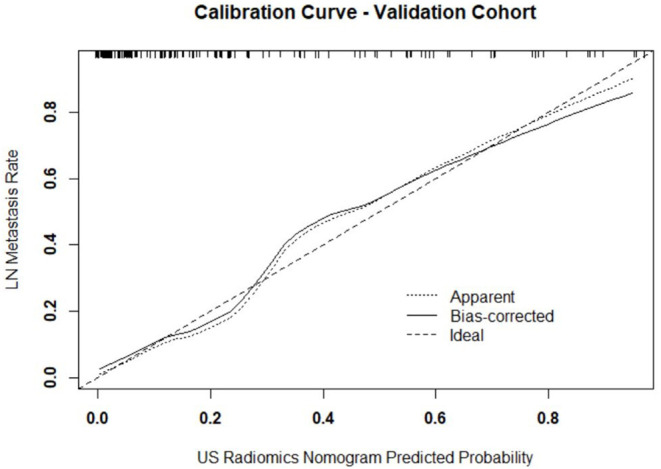
This study aimed to establish and validate an ultrasound radiomics nomogram for the preoperative prediction of central lymph node (LN) metastasis in patients with papillary thyroid carcinoma (PTC). The prediction model was developed in 609 patients with clinicopathologically confirmed unifocal PTC who received ultrasonography between Jan 2018 and June 2018. Radiomic features were extracted after the ultrasonography of PTC. Lasso regression model was used for data dimensionality reduction, feature selection, and radiomics signature building. The predicting model was established based on the multivariable logistic regression analysis in which the radiomics signature, ultrasonography-reported LN status, and independent clinicopathologic risk factors were incorporated, and finally a radiomics nomogram was established. The performance of the nomogram was assessed with respect to the discrimination and consistence. An independent validation was performed in 326 consecutive patients from July 2018 to Sep 2018. The radiomics signature consisted of 23 selected features and was significantly associated with LN status in both primary and validation cohorts. The independent predictors in the radiomics nomogram included the radiomics signature, age, TG level, TPOAB level, and ultrasonography-reported LN status. The model showed good discrimination and consistence in both cohorts: C-index of 0.816 (95% CI, 0.808-0.824) in the primary cohort and 0.858 (95% CI, 0.849-0.867) in the validation cohort. The area under receiver operating curve was 0.858. In the validation cohort, the accuracy, sensitivity, specificity and AUC of this model were 0.812, 0.816, 0.810, and 0.858 (95% CI, 0.785-0.930), respectively. Decision curve analysis indicated the radiomics nomogram was clinically useful. This study presents a convenient, clinically useful ultrasound radiomics nomogram that can be used for the pre-operative individualized prediction of central LN metastasis in patients with PTC.
本研究旨在建立并验证一种超声影像组学列线图,用于术前预测甲状腺乳头状癌(PTC)患者的中央淋巴结(LN)转移情况。该预测模型在609例经临床病理证实为单灶性PTC且于2018年1月至2018年6月期间接受超声检查的患者中构建。在对PTC进行超声检查后提取影像组学特征。采用套索回归模型进行数据降维、特征选择及影像组学特征构建。基于多变量逻辑回归分析建立预测模型,该分析纳入了影像组学特征、超声报告的LN状态及独立的临床病理危险因素,最终建立了影像组学列线图。从判别能力和一致性方面评估列线图的性能。在2018年7月至2018年9月期间连续纳入的326例患者中进行独立验证。影像组学特征由23个选定特征组成,在主要队列和验证队列中均与LN状态显著相关。影像组学列线图中的独立预测因素包括影像组学特征、年龄、TG水平、TPOAB水平及超声报告的LN状态。该模型在两个队列中均显示出良好的判别能力和一致性:主要队列中的C指数为0.816(95%CI,0.808 - 0.824),验证队列中的C指数为0.858(95%CI,0.849 - 0.867)。受试者操作特征曲线下面积为0.858。在验证队列中,该模型的准确性、敏感性、特异性及AUC分别为0.812、0.816、0.810及0.858(95%CI,0.785 - 0.930)。决策曲线分析表明影像组学列线图具有临床实用性。本研究提出了一种方便、具有临床实用性的超声影像组学列线图,可用于PTC患者术前中央LN转移的个体化预测。