• 文献检索
  • 文档翻译
  • 深度研究
  • 学术资讯
  • Suppr Zotero 插件Zotero 插件
  • 邀请有礼
  • 套餐&价格
  • 历史记录
应用&插件
Suppr Zotero 插件Zotero 插件浏览器插件Mac 客户端Windows 客户端微信小程序
定价
高级版会员购买积分包购买API积分包
服务
文献检索文档翻译深度研究API 文档MCP 服务
关于我们
关于 Suppr公司介绍联系我们用户协议隐私条款
关注我们

Suppr 超能文献

核心技术专利:CN118964589B侵权必究
粤ICP备2023148730 号-1Suppr @ 2026

文献检索

告别复杂PubMed语法,用中文像聊天一样搜索,搜遍4000万医学文献。AI智能推荐,让科研检索更轻松。

立即免费搜索

文件翻译

保留排版,准确专业,支持PDF/Word/PPT等文件格式,支持 12+语言互译。

免费翻译文档

深度研究

AI帮你快速写综述,25分钟生成高质量综述,智能提取关键信息,辅助科研写作。

立即免费体验

简要回顾一下微观研究如何阐明高等植物类囊体的 3D 结构和大分子组织。

A brief history of how microscopic studies led to the elucidation of the 3D architecture and macromolecular organization of higher plant thylakoids.

机构信息

Department of Molecular, Cellular and Developmental Biology, UCB 347, University of Colorado, Boulder, CO, 80309-0347, USA.

Plant Biology Section, School of Integrative Plant Science, Cornell University, Ithaca, NY, 14853, USA.

出版信息

Photosynth Res. 2020 Sep;145(3):237-258. doi: 10.1007/s11120-020-00782-3. Epub 2020 Oct 5.

DOI:10.1007/s11120-020-00782-3
PMID:33017036
原文链接:https://pmc.ncbi.nlm.nih.gov/articles/PMC7541383/
Abstract

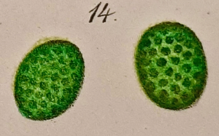
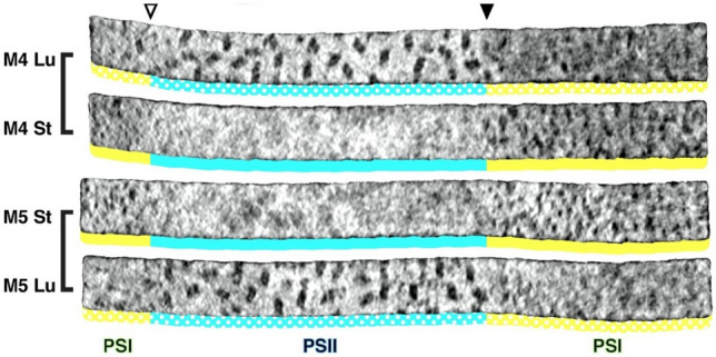
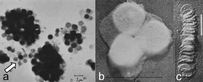
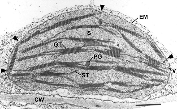
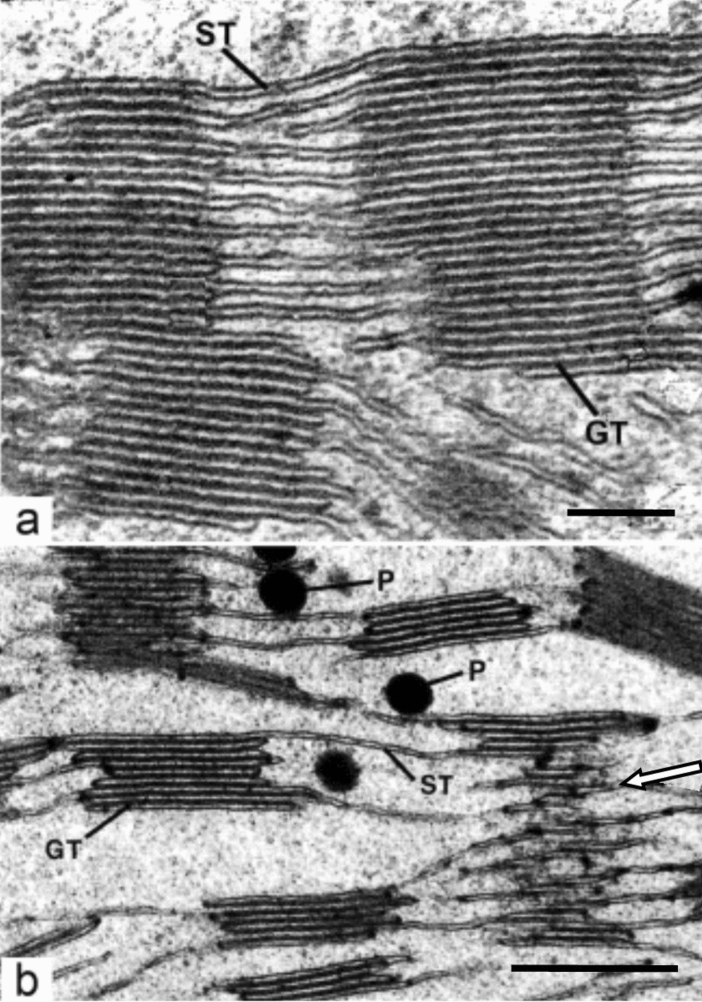
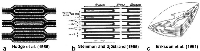
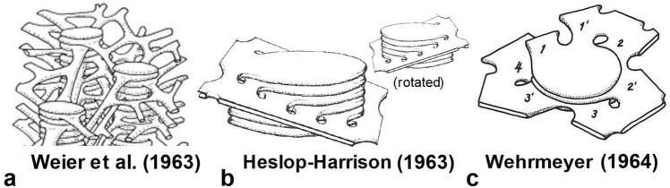
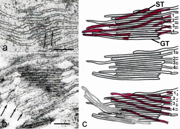
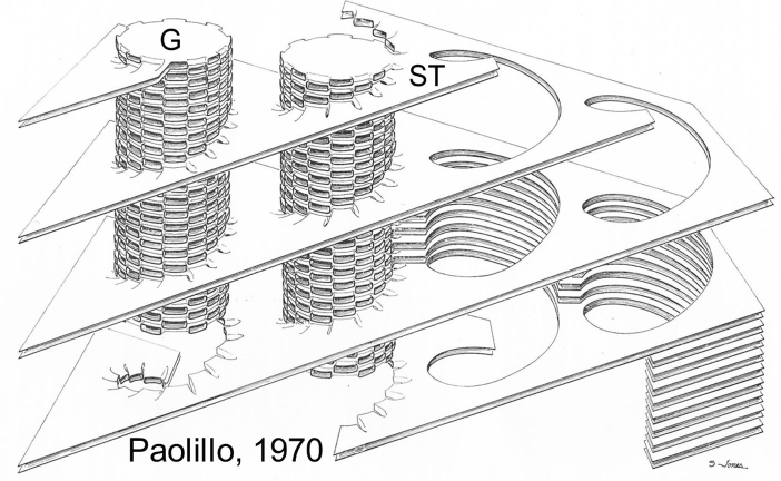
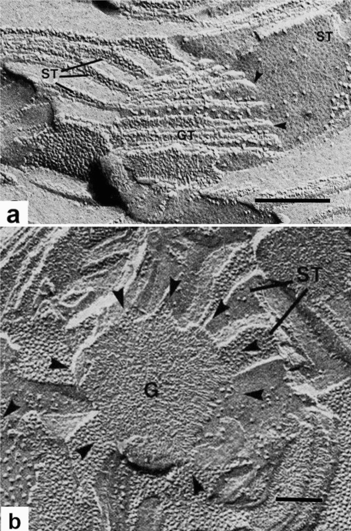
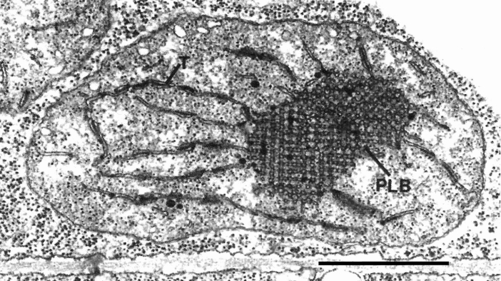
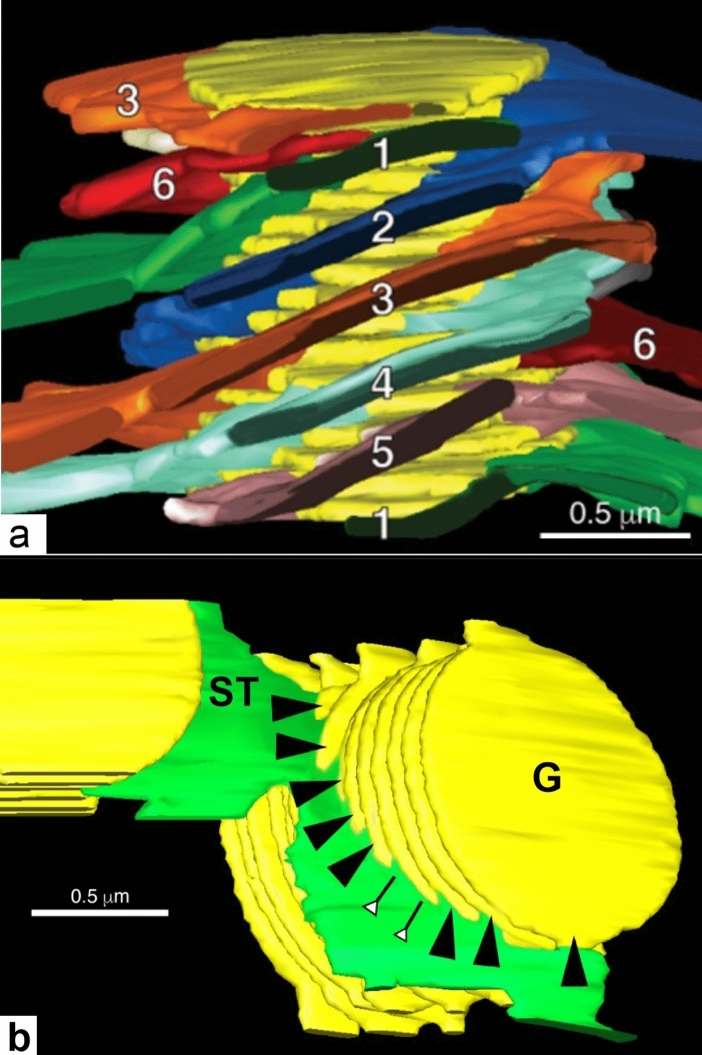
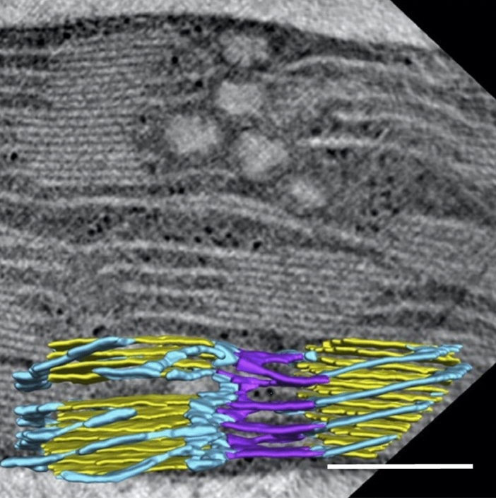
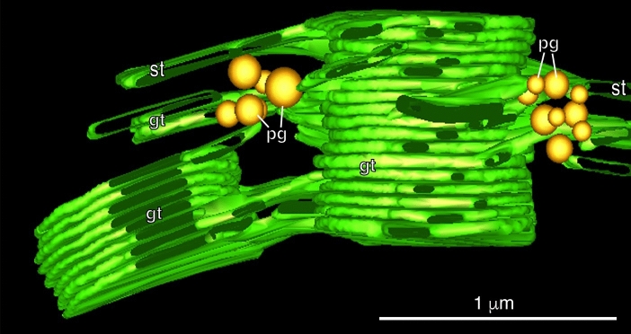
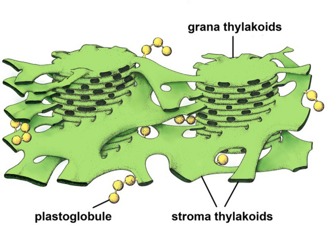
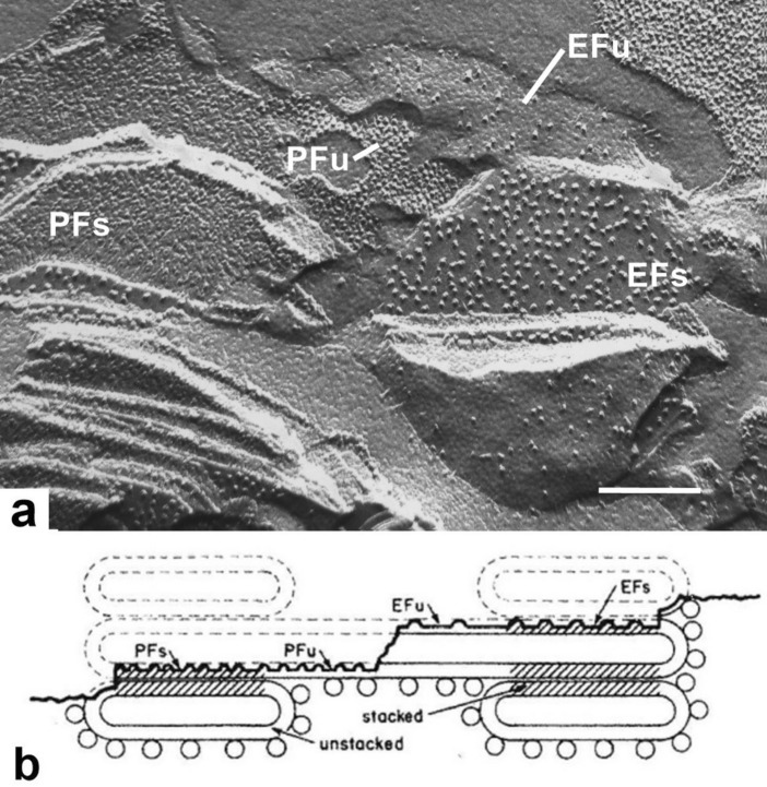
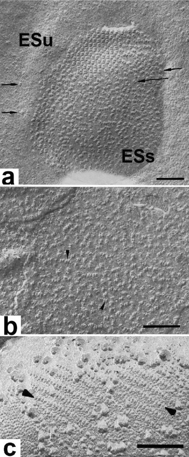
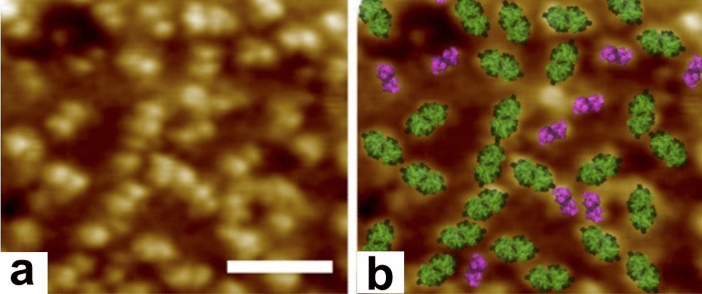
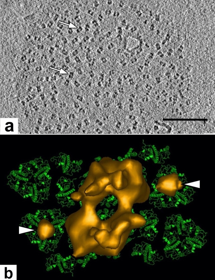
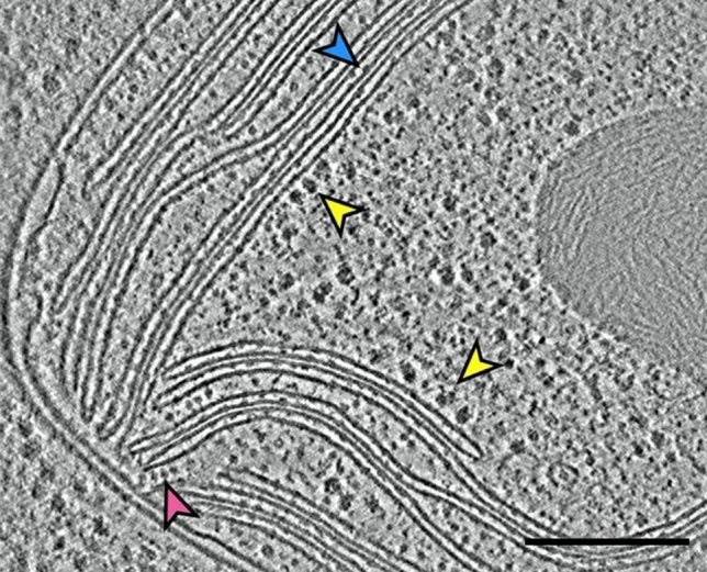
Microscopic studies of chloroplasts can be traced back to the year 1678 when Antonie van Leeuwenhoek reported to the Royal Society in London that he saw green globules in grass leaf cells with his single-lens microscope. Since then, microscopic studies have continued to contribute critical insights into the complex architecture of chloroplast membranes and how their structure relates to function. This review is organized into three chronological sections: During the classic light microscope period (1678-1940), the development of improved microscopes led to the identification of green grana, a colorless stroma, and a membrane envelope. More recent (1990-2020) chloroplast dynamic studies have benefited from laser confocal and 3D-structured illumination microscopy. The development of the transmission electron microscope (1940-2000) and thin sectioning techniques demonstrated that grana consist of stacks of closely appressed grana thylakoids interconnected by non-appressed stroma thylakoids. When the stroma thylakoids were shown to spiral around the grana stacks as multiple right-handed helices, it was confirmed that the membranes of a chloroplast are all interconnected. Freeze-fracture and freeze-etch methods verified the helical nature of the stroma thylakoids, while also providing precise information on how the electron transport chain and ATP synthase complexes are non-randomly distributed between grana and stroma membrane regions. The last section (2000-2020) focuses on the most recent discoveries made possible by atomic force microscopy of hydrated membranes, and electron tomography and cryo-electron tomography of cryofixed thylakoids. These investigations have provided novel insights into thylakoid architecture and plastoglobules (summarized in a new thylakoid model), while also producing molecular-scale views of grana and stroma thylakoids in which individual functional complexes can be identified.

摘要

叶绿体的微观研究可以追溯到 1678 年,当时安东尼·范·列文虎克(Antonie van Leeuwenhoek)向伦敦皇家学会报告说,他用单透镜显微镜在草叶细胞中看到了绿色小球。从那时起,微观研究继续为叶绿体膜的复杂结构以及其结构与功能的关系提供了关键的见解。本综述分为三个时间顺序的部分:在经典的光学显微镜时期(1678-1940 年),改进显微镜的发展导致了绿色基粒、无色基质和膜包膜的鉴定。最近(1990-2020 年)的叶绿体动态研究受益于激光共聚焦和 3D 结构照明显微镜。透射电子显微镜(1940-2000 年)和薄切片技术的发展表明,基粒由紧密贴合的基粒类囊体堆叠组成,通过非贴合的基质类囊体相互连接。当基质类囊体被证明像多个右手螺旋一样缠绕在基粒堆叠周围时,就证实了叶绿体的膜是相互连接的。冷冻断裂和冷冻蚀刻方法验证了基质类囊体的螺旋性质,同时还提供了关于电子传递链和 ATP 合酶复合物如何在基粒和基质膜区域之间非随机分布的精确信息。最后一部分(2000-2020 年)专注于原子力显微镜对水合膜、电子断层扫描和冷冻固定类囊体的电子断层扫描的最新发现。这些研究为类囊体结构和质体小球提供了新的见解(总结在一个新的类囊体模型中),同时还产生了可以识别单个功能复合物的基粒和基质类囊体的分子尺度视图。

https://cdn.ncbi.nlm.nih.gov/pmc/blobs/73f4/7541383/8c402d118437/11120_2020_782_Fig20_HTML.jpg
https://cdn.ncbi.nlm.nih.gov/pmc/blobs/73f4/7541383/1021bc4a5aea/11120_2020_782_Fig1_HTML.jpg
https://cdn.ncbi.nlm.nih.gov/pmc/blobs/73f4/7541383/15c0ae056acf/11120_2020_782_Fig2_HTML.jpg
https://cdn.ncbi.nlm.nih.gov/pmc/blobs/73f4/7541383/5d971b269eeb/11120_2020_782_Fig3_HTML.jpg
https://cdn.ncbi.nlm.nih.gov/pmc/blobs/73f4/7541383/7fec2389434a/11120_2020_782_Fig4_HTML.jpg
https://cdn.ncbi.nlm.nih.gov/pmc/blobs/73f4/7541383/c33d170adf20/11120_2020_782_Fig5_HTML.jpg
https://cdn.ncbi.nlm.nih.gov/pmc/blobs/73f4/7541383/7452407e9062/11120_2020_782_Fig6_HTML.jpg
https://cdn.ncbi.nlm.nih.gov/pmc/blobs/73f4/7541383/f9a98a570a66/11120_2020_782_Fig7_HTML.jpg
https://cdn.ncbi.nlm.nih.gov/pmc/blobs/73f4/7541383/cbe6841e998c/11120_2020_782_Fig8_HTML.jpg
https://cdn.ncbi.nlm.nih.gov/pmc/blobs/73f4/7541383/00285f9eda7a/11120_2020_782_Fig9_HTML.jpg
https://cdn.ncbi.nlm.nih.gov/pmc/blobs/73f4/7541383/b6ac75c7ac23/11120_2020_782_Fig10_HTML.jpg
https://cdn.ncbi.nlm.nih.gov/pmc/blobs/73f4/7541383/71a1a89a4b53/11120_2020_782_Fig11_HTML.jpg
https://cdn.ncbi.nlm.nih.gov/pmc/blobs/73f4/7541383/ad6618624cde/11120_2020_782_Fig12_HTML.jpg
https://cdn.ncbi.nlm.nih.gov/pmc/blobs/73f4/7541383/f0b0f81ac160/11120_2020_782_Fig13_HTML.jpg
https://cdn.ncbi.nlm.nih.gov/pmc/blobs/73f4/7541383/5357653caa03/11120_2020_782_Fig14_HTML.jpg
https://cdn.ncbi.nlm.nih.gov/pmc/blobs/73f4/7541383/927bb72aa8e7/11120_2020_782_Fig15_HTML.jpg
https://cdn.ncbi.nlm.nih.gov/pmc/blobs/73f4/7541383/14bc7511e7df/11120_2020_782_Fig16_HTML.jpg
https://cdn.ncbi.nlm.nih.gov/pmc/blobs/73f4/7541383/63073f152fbe/11120_2020_782_Fig17_HTML.jpg
https://cdn.ncbi.nlm.nih.gov/pmc/blobs/73f4/7541383/e83e4bdbc346/11120_2020_782_Fig18_HTML.jpg
https://cdn.ncbi.nlm.nih.gov/pmc/blobs/73f4/7541383/60ae2b046d74/11120_2020_782_Fig19_HTML.jpg
https://cdn.ncbi.nlm.nih.gov/pmc/blobs/73f4/7541383/8c402d118437/11120_2020_782_Fig20_HTML.jpg

相似文献

1
A brief history of how microscopic studies led to the elucidation of the 3D architecture and macromolecular organization of higher plant thylakoids.简要回顾一下微观研究如何阐明高等植物类囊体的 3D 结构和大分子组织。
Photosynth Res. 2020 Sep;145(3):237-258. doi: 10.1007/s11120-020-00782-3. Epub 2020 Oct 5.
2
Three-dimensional architecture of grana and stroma thylakoids of higher plants as determined by electron tomography.高等植物类囊体垛叠和基质类囊体的三维结构通过电子断层扫描技术确定。
Plant Physiol. 2011 Apr;155(4):1601-11. doi: 10.1104/pp.110.170647. Epub 2011 Jan 11.
3
Chloroplast structure: from chlorophyll granules to supra-molecular architecture of thylakoid membranes.叶绿体结构:从叶绿素颗粒到类囊体膜的超分子结构
Photosynth Res. 2003;76(1-3):185-96. doi: 10.1023/A:1024994525586.
4
Dynamic flexibility in the structure and function of photosystem II in higher plant thylakoid membranes: the grana enigma.高等植物类囊体膜中光系统II结构与功能的动态灵活性:基粒之谜
Photosynth Res. 2008 Oct-Dec;98(1-3):575-87. doi: 10.1007/s11120-008-9381-3. Epub 2008 Nov 8.
5
Insights into the complex 3-D architecture of thylakoid membranes in unicellular cyanobacterium Cyanothece sp. ATCC 51142.揭示单细胞蓝藻集胞藻 Cyanothece sp. ATCC 51142 中类囊体膜的复杂三维结构。
Plant Signal Behav. 2011 Apr;6(4):566-9. doi: 10.4161/psb.6.4.14946. Epub 2011 Apr 1.
6
Structure and dynamics of thylakoids in land plants.类囊体在陆生植物中的结构与动态。
J Exp Bot. 2014 May;65(8):1955-72. doi: 10.1093/jxb/eru090. Epub 2014 Mar 12.
7
Thylakoid-Bound Polysomes and a Dynamin-Related Protein, FZL, Mediate Critical Stages of the Linear Chloroplast Biogenesis Program in Greening Arabidopsis Cotyledons.类囊体结合多核糖体和一个与动力蛋白相关的蛋白 FZL,在拟南芥子叶的线性叶绿体生物发生程序的关键阶段起作用。
Plant Cell. 2018 Jul;30(7):1476-1495. doi: 10.1105/tpc.17.00972. Epub 2018 Jun 7.
8
Electron tomography of plant thylakoid membranes.植物类囊体膜的电子断层扫描。
J Exp Bot. 2011 Apr;62(7):2393-402. doi: 10.1093/jxb/err034. Epub 2011 Mar 25.
9
Expansion microscopy resolves the thylakoid structure of spinach.扩展显微镜解析菠菜类囊体结构。
Plant Physiol. 2023 Dec 30;194(1):347-358. doi: 10.1093/plphys/kiad526.
10
An alternative model for photosystem II/light harvesting complex II in grana membranes based on cryo-electron microscopy studies.基于冷冻电子显微镜研究的叶绿体基粒膜中光系统II/捕光复合物II的替代模型。
Eur J Biochem. 2002 Jan;269(1):326-36. doi: 10.1046/j.0014-2956.2001.02652.x.

引用本文的文献

1
GRANA: An AI-based tool for accelerating chloroplast grana nanomorphology analysis using hybrid intelligence.GRANA:一种基于人工智能的工具,用于利用混合智能加速叶绿体基粒纳米形态分析。
Plant Physiol. 2025 May 30;198(2). doi: 10.1093/plphys/kiaf212.
2
Cryo-ET reveals the in situ architecture of the polar tube invasion apparatus from microsporidian parasites.冷冻电子断层扫描揭示了微孢子虫寄生虫极管侵入装置的原位结构。
Proc Natl Acad Sci U S A. 2025 Mar 18;122(11):e2415233122. doi: 10.1073/pnas.2415233122. Epub 2025 Mar 11.
3
Comparative Insights into Photosynthetic, Biochemical, and Ultrastructural Mechanisms in Hibiscus and Pelargonium Plants.

本文引用的文献

1
Charting the native architecture of thylakoid membranes with single-molecule precision.用单分子精度绘制类囊体膜的天然结构。
Elife. 2020 Apr 16;9:e53740. doi: 10.7554/eLife.53740.
2
A Sec14 domain protein is required for photoautotrophic growth and chloroplast vesicle formation in .Sec14 结构域蛋白是莱茵衣藻光自养生长和叶绿体囊泡形成所必需的。
Proc Natl Acad Sci U S A. 2020 Apr 21;117(16):9101-9111. doi: 10.1073/pnas.1916946117. Epub 2020 Apr 3.
3
Fundamental helical geometry consolidates the plant photosynthetic membrane.
木槿和天竺葵植物光合作用、生化及超微结构机制的比较研究
Plants (Basel). 2024 Oct 9;13(19):2831. doi: 10.3390/plants13192831.
4
Chloroplast biogenesis involves spatial coordination of nuclear and organellar gene expression in Chlamydomonas.叶绿体生物发生涉及衣藻中核和细胞器基因表达的空间协调。
Plant Physiol. 2024 Sep 2;196(1):112-123. doi: 10.1093/plphys/kiae256.
5
Thylakoid ultrastructural variations in chlorophyll-deficient wheat: aberrations or structural acclimation?类囊体超微结构在缺叶绿素小麦中的变化:是畸变还是结构适应?
Planta. 2024 Mar 13;259(4):90. doi: 10.1007/s00425-024-04362-w.
6
Eukaryotic Cell Membranes: Structure, Composition, Research Methods and Computational Modelling.真核细胞的膜结构、组成、研究方法和计算建模。
Int J Mol Sci. 2023 Jul 7;24(13):11226. doi: 10.3390/ijms241311226.
7
Light energy transduction in liposome-based artificial cells.基于脂质体的人工细胞中的光能转导。
Front Bioeng Biotechnol. 2023 Mar 29;11:1161730. doi: 10.3389/fbioe.2023.1161730. eCollection 2023.
8
From Protein Film Electrochemistry to Nanoconfined Enzyme Cascades and the Electrochemical Leaf.从蛋白质膜电化学到纳米受限酶级联反应和电化学叶。
Chem Rev. 2023 May 10;123(9):5421-5458. doi: 10.1021/acs.chemrev.2c00397. Epub 2022 Dec 27.
9
State transition is quiet around pyrenoid and LHCII phosphorylation is not essential for thylakoid deformation in 137c.在 137c 中,淀粉核周围的状态转换是安静的,类囊体膜上 LHCII 的磷酸化对于类囊体膜的变形不是必需的。
Proc Natl Acad Sci U S A. 2022 Sep 13;119(37):e2122032119. doi: 10.1073/pnas.2122032119. Epub 2022 Sep 6.
10
Electron microscopy for imaging organelles in plants and algae.用于成像植物和藻类细胞器的电子显微镜。
Plant Physiol. 2022 Feb 4;188(2):713-725. doi: 10.1093/plphys/kiab449.
基础螺旋几何结构巩固了植物光合作用膜。
Proc Natl Acad Sci U S A. 2019 Oct 29;116(44):22366-22375. doi: 10.1073/pnas.1905994116. Epub 2019 Oct 14.
4
A brief history of thylakoid biogenesis.类囊体生物发生简史。
Open Biol. 2019 Jan 31;9(1):180237. doi: 10.1098/rsob.180237.
5
Chloroplast vesicle transport.叶绿体小泡运输。
Photosynth Res. 2018 Dec;138(3):361-371. doi: 10.1007/s11120-018-0566-0. Epub 2018 Aug 16.
6
Subdiffraction-resolution live-cell imaging for visualizing thylakoid membranes.亚衍射分辨率活细胞成像,用于可视化类囊体膜。
Plant J. 2018 Oct;96(1):233-243. doi: 10.1111/tpj.14021. Epub 2018 Jul 30.
7
Thylakoid-Bound Polysomes and a Dynamin-Related Protein, FZL, Mediate Critical Stages of the Linear Chloroplast Biogenesis Program in Greening Arabidopsis Cotyledons.类囊体结合多核糖体和一个与动力蛋白相关的蛋白 FZL,在拟南芥子叶的线性叶绿体生物发生程序的关键阶段起作用。
Plant Cell. 2018 Jul;30(7):1476-1495. doi: 10.1105/tpc.17.00972. Epub 2018 Jun 7.
8
Dynamic thylakoid stacking regulates the balance between linear and cyclic photosynthetic electron transfer.动态类囊体垛叠调节线性和循环光合电子传递之间的平衡。
Nat Plants. 2018 Feb;4(2):116-127. doi: 10.1038/s41477-017-0092-7. Epub 2018 Jan 29.
9
Plastoglobuli: Plastid Microcompartments with Integrated Functions in Metabolism, Plastid Developmental Transitions, and Environmental Adaptation.质体小球体:代谢、质体发育转变和环境适应中具有集成功能的质体微区室。
Annu Rev Plant Biol. 2017 Apr 28;68:253-289. doi: 10.1146/annurev-arplant-043015-111737. Epub 2017 Jan 25.
10
Three-Dimensional Visualization of the Tubular-Lamellar Transformation of the Internal Plastid Membrane Network during Runner Bean Chloroplast Biogenesis.菜豆叶绿体生物发生过程中质体内膜网络管状-片层转变的三维可视化
Plant Cell. 2016 Apr;28(4):875-91. doi: 10.1105/tpc.15.01053. Epub 2016 Mar 21.