• 文献检索
  • 文档翻译
  • 深度研究
  • 学术资讯
  • Suppr Zotero 插件Zotero 插件
  • 邀请有礼
  • 套餐&价格
  • 历史记录
应用&插件
Suppr Zotero 插件Zotero 插件浏览器插件Mac 客户端Windows 客户端微信小程序
定价
高级版会员购买积分包购买API积分包
服务
文献检索文档翻译深度研究API 文档MCP 服务
关于我们
关于 Suppr公司介绍联系我们用户协议隐私条款
关注我们

Suppr 超能文献

核心技术专利:CN118964589B侵权必究
粤ICP备2023148730 号-1Suppr @ 2026

文献检索

告别复杂PubMed语法,用中文像聊天一样搜索,搜遍4000万医学文献。AI智能推荐,让科研检索更轻松。

立即免费搜索

文件翻译

保留排版,准确专业,支持PDF/Word/PPT等文件格式,支持 12+语言互译。

免费翻译文档

深度研究

AI帮你快速写综述,25分钟生成高质量综述,智能提取关键信息,辅助科研写作。

立即免费体验

原发性开角型青光眼结构连接组的重组织。

Reorganization of the structural connectome in primary open angle Glaucoma.

机构信息

Medical Physics Section, Department of Biomedicine and Prevention, University of Rome "Tor Vergata", Italy.

Neuroradiology Unit, Department of Biomedicine and Prevention, University of Rome "Tor Vergata", Rome, Italy; San Raffaele Cassino, Frosinone, Italy.

出版信息

Neuroimage Clin. 2020;28:102419. doi: 10.1016/j.nicl.2020.102419. Epub 2020 Sep 9.

DOI:10.1016/j.nicl.2020.102419
PMID:33032067
原文链接:https://pmc.ncbi.nlm.nih.gov/articles/PMC7552094/
Abstract

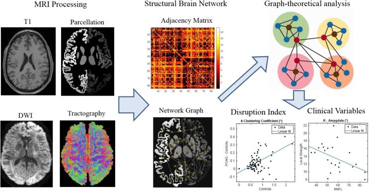
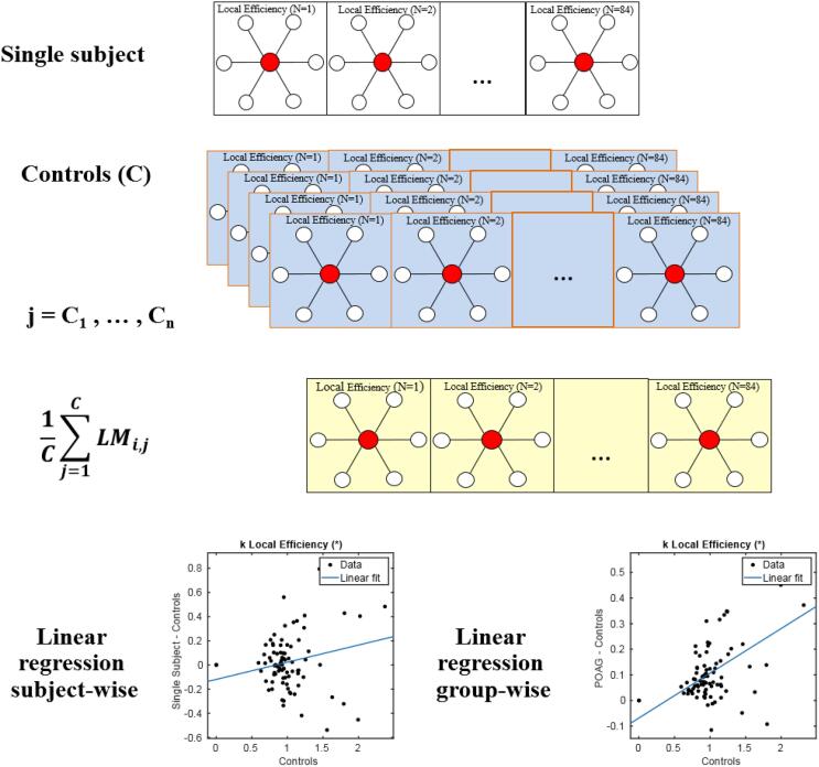
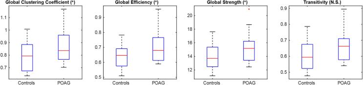
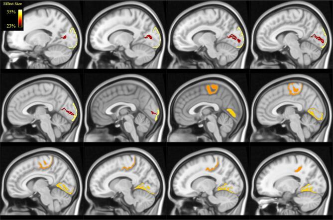
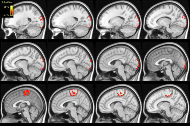
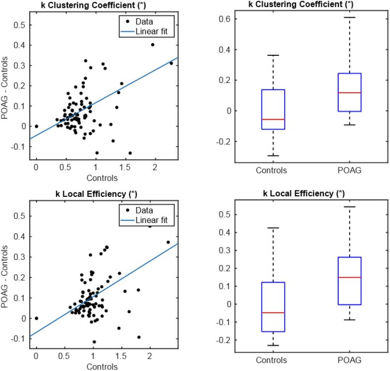
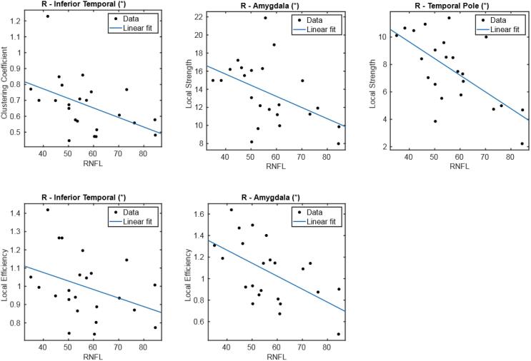
Primary open angle Glaucoma (POAG) is one of the most common causes of permanent blindness in the world. Recent studies have suggested the hypothesis that POAG is also a central nervous system disorder which may result in additional (i.e., extra-ocular) involvement. The aim of this study is to assess possible structural, whole-brain connectivity alterations in POAG patients. We evaluated 23 POAG patients and 15 healthy controls by combining multi-shell diffusion weighted imaging, multi-shell, multi-tissue probabilistic tractography, graph theoretical measures and a recently designed 'disruption index', which evaluates the global reorganization of brain networks. We also studied the associations between the whole-brain structural connectivity measures and indices of visual acuity including the field index (VFI) and two Optical Coherence Tomography (OCT) parameters, namely the Macula Ganglion Cell Layer (MaculaGCL) and Retinal Nerve Fiber Layer (RNFL) thicknesses. We found both global and local structural connectivity differences between POAG patients and controls, which extended well beyond the primary visual pathway and were localized in the left calcarine gyrus (clustering coefficient p = 0.036), left lateral occipital cortex (clustering coefficient p = 0.017, local efficiency p = 0.035), right lingual gyrus (clustering coefficient p = 0.009), and right paracentral lobule (clustering coefficient p = 0.009, local efficiency p = 0.018). Group-wise (clustering coefficient, p = 6.59∙10 and local efficiency p = 6.23·10) and subject-wise disruption indices (clustering coefficient, p = 0.018 and local efficiency, p = 0.01) also differed between POAG patients and controls. In addition, we found negative associations between RNFL thickness and local measures (clustering coefficient, local efficiency and strength) in the right amygdala (local efficiency p = 0.008, local strength p = 0.016), right inferior temporal gyrus (clustering coefficient p = 0.036, local efficiency p = 0.042), and right temporal pole (local strength p = 0.008). Overall, we show, in patients with POAG, a whole-brain structural reorganization that spans across a variety of brain regions involved in visual processing, motor control, and emotional/cognitive functions. We also identified a pattern of brain structural changes in relation to POAG clinical severity. Taken together, our findings support the hypothesis that the reduction in visual acuity from POAG can be driven by a combination of local (i.e., in the eye) and more extended (i.e., brain) effects.

摘要

原发性开角型青光眼(POAG)是世界上导致永久性失明的最常见原因之一。最近的研究表明,POAG 也是一种中枢神经系统疾病,可能导致额外的(即眼外)受累。本研究旨在评估 POAG 患者可能存在的结构和全脑连接改变。我们通过结合多壳扩散加权成像、多壳、多组织概率追踪、图论测量和最近设计的“破坏指数”,评估了 23 名 POAG 患者和 15 名健康对照者的结构。我们还研究了全脑结构连接测量值与视力指标(包括视野指数(VFI)和两种光学相干断层扫描(OCT)参数,即黄斑神经节细胞层(MaculaGCL)和视网膜神经纤维层(RNFL)厚度)之间的相关性。我们发现 POAG 患者和对照组之间存在全脑和局部结构连接差异,这些差异不仅延伸到初级视觉通路,而且还局限于左侧距状回(聚类系数 p=0.036)、左侧外侧枕叶皮层(聚类系数 p=0.017,局部效率 p=0.035)、右侧舌回(聚类系数 p=0.009)和右侧中央旁小叶(聚类系数 p=0.009,局部效率 p=0.018)。组间(聚类系数,p=6.59·10 和局部效率,p=6.23·10)和个体间破坏指数(聚类系数,p=0.018 和局部效率,p=0.01)也存在差异。此外,我们还发现右侧杏仁核(局部效率,p=0.018,局部强度,p=0.01)的局部测量值(聚类系数、局部效率和强度)与 RNFL 厚度之间存在负相关,右侧颞下回(聚类系数 p=0.036,局部效率 p=0.042)和右侧颞极(局部强度 p=0.008)。总的来说,我们在 POAG 患者中发现了一种跨越多种参与视觉处理、运动控制和情感/认知功能的脑区的全脑结构重排。我们还确定了与 POAG 临床严重程度相关的脑结构变化模式。总之,我们的研究结果支持这样的假设,即 POAG 导致的视力下降可能是由局部(即眼内)和更广泛(即大脑)效应共同作用的结果。

https://cdn.ncbi.nlm.nih.gov/pmc/blobs/7d9e/7552094/bad76f63e24a/gr9.jpg
https://cdn.ncbi.nlm.nih.gov/pmc/blobs/7d9e/7552094/89bcd9981fe2/gr1.jpg
https://cdn.ncbi.nlm.nih.gov/pmc/blobs/7d9e/7552094/377eb6dc3e47/gr2.jpg
https://cdn.ncbi.nlm.nih.gov/pmc/blobs/7d9e/7552094/31882f5686f3/gr3.jpg
https://cdn.ncbi.nlm.nih.gov/pmc/blobs/7d9e/7552094/b44a2c369773/gr4.jpg
https://cdn.ncbi.nlm.nih.gov/pmc/blobs/7d9e/7552094/f2083ea60a70/gr5.jpg
https://cdn.ncbi.nlm.nih.gov/pmc/blobs/7d9e/7552094/9560dd95d491/gr6.jpg
https://cdn.ncbi.nlm.nih.gov/pmc/blobs/7d9e/7552094/57265ea77731/gr7.jpg
https://cdn.ncbi.nlm.nih.gov/pmc/blobs/7d9e/7552094/177ec5ed0029/gr8.jpg
https://cdn.ncbi.nlm.nih.gov/pmc/blobs/7d9e/7552094/bad76f63e24a/gr9.jpg

相似文献

1
Reorganization of the structural connectome in primary open angle Glaucoma.原发性开角型青光眼结构连接组的重组织。
Neuroimage Clin. 2020;28:102419. doi: 10.1016/j.nicl.2020.102419. Epub 2020 Sep 9.
2
Disruption of structural brain networks in Primary Open Angle Glaucoma.原发性开角型青光眼患者脑结构网络的破坏
Annu Int Conf IEEE Eng Med Biol Soc. 2020 Jul;2020:1705-1708. doi: 10.1109/EMBC44109.2020.9175417.
3
Primary Open Angle Glaucoma Is Associated With Functional Brain Network Reorganization.原发性开角型青光眼与功能性脑网络重组有关。
Front Neurol. 2019 Oct 25;10:1134. doi: 10.3389/fneur.2019.01134. eCollection 2019.
4
Structural brain abnormalities in patients with primary open-angle glaucoma: a study with 3T MR imaging.原发性开角型青光眼患者的脑结构异常:一项 3T MRI 研究。
Invest Ophthalmol Vis Sci. 2013 Jan 17;54(1):545-54. doi: 10.1167/iovs.12-9893.
5
Early changes of brain connectivity in primary open angle glaucoma.原发性开角型青光眼患者脑连接的早期变化
Hum Brain Mapp. 2016 Dec;37(12):4581-4596. doi: 10.1002/hbm.23330. Epub 2016 Aug 9.
6
[The function-structure impairment pattern of optic nerves in primary open-angle glaucoma and normal-tension glaucoma].[原发性开角型青光眼和正常眼压性青光眼视神经的功能-结构损害模式]
Zhonghua Yan Ke Za Zhi. 2018 Nov 11;54(11):811-819. doi: 10.3760/cma.j.issn.0412-4081.2018.11.004.
7
[Analysis of morphologic changes of lamina cribrosa in primary open angle glaucoma using enhanced depth imaging optical coherence tomography].[使用增强深度成像光学相干断层扫描分析原发性开角型青光眼筛板的形态学变化]
Zhonghua Yan Ke Za Zhi. 2016 Jun 11;52(6):422-8. doi: 10.3760/cma.j.issn.0412-4081.2016.06.006.
8
Intraeye retinal nerve fiber layer and macular thickness asymmetry measurements for the discrimination of primary open-angle glaucoma and normal tension glaucoma.用于鉴别原发性开角型青光眼和正常眼压性青光眼的眼内视网膜神经纤维层及黄斑厚度不对称测量
J Optom. 2016 Apr-Jun;9(2):118-25. doi: 10.1016/j.optom.2015.10.002. Epub 2015 Dec 1.
9
Data-Driven Parcellation Approaches Based on Functional Connectivity of Visual Cortices in Primary Open-Angle Glaucoma.基于原发性开角型青光眼视皮层功能连接的基于数据驱动的脑区划分方法。
Invest Ophthalmol Vis Sci. 2020 Jul 1;61(8):33. doi: 10.1167/iovs.61.8.33.
10
Comparison of ganglion cell-inner plexiform layer thickness in exfoliative glaucoma and primary open-angle glaucoma.原发性开角型青光眼和剥脱性青光眼的神经节细胞-内丛状层厚度比较。
Photodiagnosis Photodyn Ther. 2021 Jun;34:102335. doi: 10.1016/j.pdpdt.2021.102335. Epub 2021 May 8.

引用本文的文献

1
Associations of diabetes mellitus with primary open angle glaucoma and Alzheimer's disease: a large cohort study in UK biobank.糖尿病与原发性开角型青光眼及阿尔茨海默病的关联:英国生物银行的一项大型队列研究
Front Endocrinol (Lausanne). 2025 Jul 24;16:1506560. doi: 10.3389/fendo.2025.1506560. eCollection 2025.
2
Abnormal topological properties and functional-structural coupling of large-scale brain networks in primary angle-closure glaucoma.原发性闭角型青光眼患者大脑大规模网络的异常拓扑特性及功能-结构耦合
Quant Imaging Med Surg. 2025 Jul 1;15(7):6053-6067. doi: 10.21037/qims-24-1947. Epub 2025 Jun 30.
3
Application of advanced diffusion MRI based tractometry of the visual pathway in glaucoma: a systematic review.

本文引用的文献

1
Variability and Reproducibility of Directed and Undirected Functional MRI Connectomes in the Human Brain.人类大脑中定向和非定向功能磁共振成像连接组的变异性和可重复性
Entropy (Basel). 2019 Jul 6;21(7):661. doi: 10.3390/e21070661.
2
Altered Large-Scale Brain Functional Connectivity in Ocular Hypertension.高眼压症患者大脑大规模功能连接的改变
Front Neurosci. 2020 Mar 5;14:146. doi: 10.3389/fnins.2020.00146. eCollection 2020.
3
Disruption of brain network organization in primary open angle glaucoma.原发性开角型青光眼中脑网络组织的破坏
基于先进扩散磁共振成像的视路纤维束成像技术在青光眼诊断中的应用:一项系统评价
Front Neurosci. 2025 May 21;19:1577991. doi: 10.3389/fnins.2025.1577991. eCollection 2025.
4
Association Between Glaucoma and Brain Structural Connectivity Based on Diffusion Tensor Tractography: A Bidirectional Mendelian Randomization Study.基于扩散张量纤维束成像的青光眼与脑结构连通性之间的关联:一项双向孟德尔随机化研究
Brain Sci. 2024 Oct 17;14(10):1030. doi: 10.3390/brainsci14101030.
5
Quality of life and mental health status of glaucoma patients.青光眼患者的生活质量和心理健康状况
Front Med (Lausanne). 2024 Jun 3;11:1402604. doi: 10.3389/fmed.2024.1402604. eCollection 2024.
6
Topological Organization of the Brain Network in Patients with Primary Angle-closure Glaucoma Through Graph Theory Analysis.通过图论分析原发性闭角型青光眼患者的大脑网络拓扑组织。
Brain Topogr. 2024 Nov;37(6):1171-1185. doi: 10.1007/s10548-024-01060-4. Epub 2024 Jun 1.
7
Diffusion tensor imaging of optic neuropathies: a narrative review.视神经病变的扩散张量成像:一项叙述性综述。
Quant Imaging Med Surg. 2024 Jan 3;14(1):1086-1107. doi: 10.21037/qims-23-779. Epub 2024 Jan 2.
8
Arterial spin labeling reveals disordered cerebral perfusion and cerebral blood flow-based functional connectivity in primary open-angle glaucoma.动脉自旋标记显示原发性开角型青光眼中存在紊乱的脑灌注和基于脑血流的功能连接。
Brain Imaging Behav. 2024 Feb;18(1):231-242. doi: 10.1007/s11682-023-00813-2. Epub 2023 Nov 25.
9
Abnormal Topological Organization of White Matter Structural Networks in Normal Tension Glaucoma Revealed via Diffusion Tensor Tractography.通过扩散张量纤维束成像揭示正常眼压性青光眼中白质结构网络的异常拓扑组织
Brain Sci. 2023 Nov 17;13(11):1597. doi: 10.3390/brainsci13111597.
10
MRI and Clinical Biomarkers Overlap between Glaucoma and Alzheimer's Disease.MRI 和临床生物标志物在青光眼和阿尔茨海默病之间存在重叠。
Int J Mol Sci. 2023 Oct 5;24(19):14932. doi: 10.3390/ijms241914932.
Annu Int Conf IEEE Eng Med Biol Soc. 2019 Jul;2019:4338-4341. doi: 10.1109/EMBC.2019.8857290.
4
Brain morphological alterations of cerebral cortex and subcortical nuclei in high-tension glaucoma brain and its associations with intraocular pressure.脑高压性青光眼患者大脑皮质和皮质下核的形态改变及其与眼内压的关系。
Neuroradiology. 2020 Apr;62(4):495-502. doi: 10.1007/s00234-019-02347-1. Epub 2019 Dec 23.
5
Primary Open Angle Glaucoma Is Associated With Functional Brain Network Reorganization.原发性开角型青光眼与功能性脑网络重组有关。
Front Neurol. 2019 Oct 25;10:1134. doi: 10.3389/fneur.2019.01134. eCollection 2019.
6
Widespread brain reorganization perturbs visuomotor coordination in early glaucoma.广泛的大脑重组扰乱了早期青光眼的视动协调。
Sci Rep. 2019 Oct 2;9(1):14168. doi: 10.1038/s41598-019-50793-x.
7
Fixel-Based Analysis of Visual Pathway White Matter in Primary Open-Angle Glaucoma.基于体素的原发性开角型青光眼视路白质分析。
Invest Ophthalmol Vis Sci. 2019 Sep 3;60(12):3803-3812. doi: 10.1167/iovs.19-27447.
8
MRtrix3: A fast, flexible and open software framework for medical image processing and visualisation.MRtrix3:一个用于医学图像处理和可视化的快速、灵活、开放的软件框架。
Neuroimage. 2019 Nov 15;202:116137. doi: 10.1016/j.neuroimage.2019.116137. Epub 2019 Aug 29.
9
Altered functional connectivity density in primary angle-closure glaucoma patients at resting-state.原发性闭角型青光眼患者静息状态下功能连接密度的改变
Quant Imaging Med Surg. 2019 Apr;9(4):603-614. doi: 10.21037/qims.2019.04.13.
10
Structural connectivity-based topography of the human globus pallidus: Implications for therapeutic targeting in movement disorders.基于结构连接的人类苍白球拓扑:在运动障碍中的治疗靶点的启示。
Mov Disord. 2019 Jul;34(7):987-996. doi: 10.1002/mds.27712. Epub 2019 May 11.