Lozar Taja, Jesenko Tanja, Kloboves Prevodnik Veronika, Cemazar Maja, Hosta Violeta, Jericevic Anja, Nolde Natasa, Grasic Kuhar Cvetka
Faculty of Medicine, University of Ljubljana, Ljubljana, Slovenia.
Department of Experimental Oncology, Institute of Oncology Ljubljana, Ljubljana, Slovenia.
Front Oncol. 2020 Sep 15;10:554554. doi: 10.3389/fonc.2020.554554. eCollection 2020.
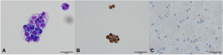
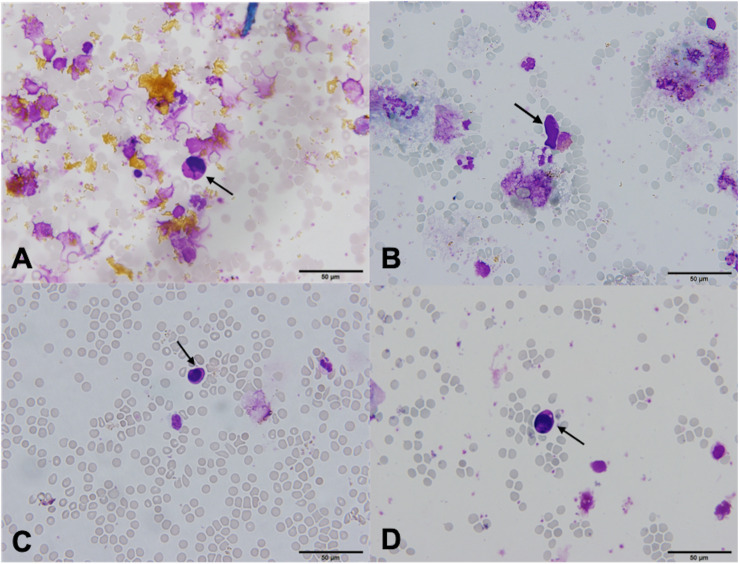
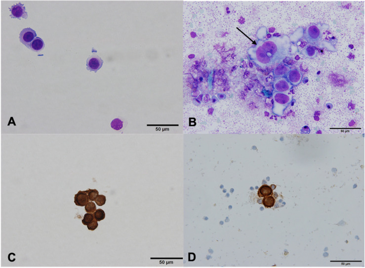
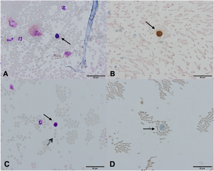
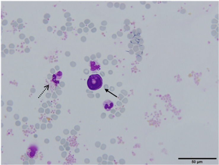
Circulating tumor cell (CTC) count is an independent prognostic factor in early breast cancer. CTCs can be found in the blood of 20% of patients prior to neoadjuvant therapy. We aimed to assess the suitability of magnetic-activated cell separation (MACS) technology for isolation and cytological characterization of CTCs. In the preclinical part of the study, cell lines were spiked into buffy coat samples derived from healthy donors, and isolated using MACS. Breast cancer cells with preserved cell morphology were successfully isolated. In the clinical part, blood for CTC isolation was drawn from 44 patients with early and locally advanced breast cancer prior to neoadjuvant chemotherapy. Standard Giemsa, Papanicolaou and pancytokeratin staining was applied. 2.3% of samples contained cells that meet both the morphological and immunocytochemical criteria for CTC. In 32.6% of samples, partially degenerated pancytokeratin negative cells with morphological features of tumor cells were observed. In 65.1% of samples, CTCs were not found. In conclusion, our results demonstrate that morphologically intact tumor cells can be isolated using MACS technology. However, morphologically intact tumor cells were not detected in the clinical part of the study. At present, MACS technology does not appear suitable for use in a clinical cytopathology laboratory.
循环肿瘤细胞(CTC)计数是早期乳腺癌的一个独立预后因素。在新辅助治疗前,20%的患者血液中可检测到循环肿瘤细胞。我们旨在评估磁激活细胞分选(MACS)技术用于循环肿瘤细胞分离及细胞学特征分析的适用性。在该研究的临床前部分,将细胞系加入来自健康供体的血沉棕黄层样本中,并用MACS进行分离。成功分离出细胞形态保持完整的乳腺癌细胞。在临床部分,于新辅助化疗前从44例早期及局部晚期乳腺癌患者抽取用于循环肿瘤细胞分离的血液。采用标准吉姆萨染色、巴氏染色和全细胞角蛋白染色。2.3%的样本中含有符合循环肿瘤细胞形态学和免疫细胞化学标准的细胞。在32.6%的样本中,观察到具有肿瘤细胞形态特征的部分退化的全细胞角蛋白阴性细胞。在65.1%的样本中未发现循环肿瘤细胞。总之,我们的结果表明,可使用MACS技术分离形态完整的肿瘤细胞。然而,在该研究的临床部分未检测到形态完整的肿瘤细胞。目前,MACS技术似乎不适用于临床细胞病理学实验室。