Karakaya Mustafa, Demirbaş Ahmet Emin
Sancaktepe Oral and Dental Health Hospital, Department of Oral and Maxillofacial Surgery, Ministry of Health, İstanbul, Turkey.
Department of Oral and Maxillofacial Surgery, Erciyes University Faculty of Dentistry, Melikgazi, Kayseri, Turkey.
Int J Implant Dent. 2020 Oct 12;6(1):61. doi: 10.1186/s40729-020-00257-z.
The primary aim of this study is to assess, in an animal model, whether biostimulation of osteoporotic bone with low-level laser therapy improves the osseointegration of dental implants.
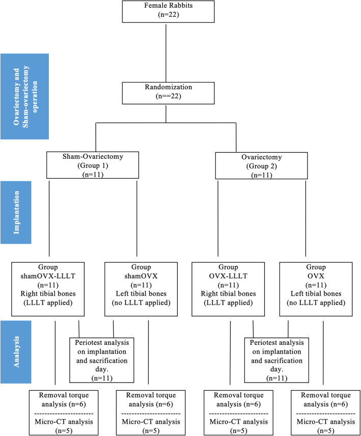
Twenty-two female rabbits were randomly divided into two groups: sham-ovariectomy and bilateral-ovariectomy. Laser therapy was applied to the implants placed in the right tibial bones and was not applied to implants placed in the left tibial bones. The periotest device was used for the stability test. Periotest values were recorded after the implantation (T0) and when the animals were euthanized (T1). The removal torque test and micro-computed tomography examination were evaluated.
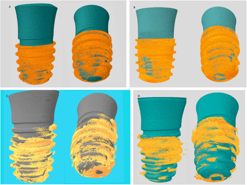
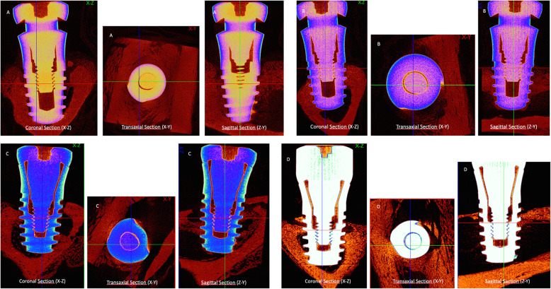
As a result of removal torque, the mean of ovariectomy-laser group (56.1 ± 5.1 Ncm) was higher than sham-ovariectomy group (55.4 ± 18.5 Ncm) (p = 0.9). In periotest analysis, a significant difference was found between the values of T1 and T0 in all groups, except sham-ovariectomy group (p < 0.05); and the highest difference was found in the ovariectomy-laser group. Micro-CT examination demonstrated that ovariectomy-laser group showed an increase of implant-bone contact when compared with ovariectomy (p < 0.05).
The values obtained from biomechanical tests and micro-CT in the ovariectomy-laser group were significantly higher than the ovariectomy group and achieved the values in the healthy bone.
本研究的主要目的是在动物模型中评估低强度激光疗法对骨质疏松骨的生物刺激是否能改善牙种植体的骨整合。
22只雌性兔子被随机分为两组:假卵巢切除组和双侧卵巢切除组。对植入右胫骨的种植体进行激光治疗,而对植入左胫骨的种植体不进行激光治疗。使用牙周测试仪进行稳定性测试。在植入后(T0)以及动物安乐死时(T1)记录牙周测试值。评估了移除扭矩测试和微型计算机断层扫描检查。
在移除扭矩测试中,卵巢切除 - 激光组的平均值(56.1 ± 5.1 Ncm)高于假卵巢切除组(55.4 ± 18.5 Ncm)(p = 0.9)。在牙周测试分析中,除假卵巢切除组外,所有组的T1和T0值之间均存在显著差异(p < 0.05);且卵巢切除 - 激光组的差异最大。微型计算机断层扫描检查表明,与卵巢切除组相比,卵巢切除 - 激光组的种植体与骨的接触增加(p < 0.05)。
卵巢切除 - 激光组从生物力学测试和微型计算机断层扫描获得的值显著高于卵巢切除组,并达到了健康骨中的值。