Division of Cardiovascular Sciences University of Manchester United Kingdom.
Department of Human and Animal Physiology Lomonosov Moscow State University Moscow Russia.
J Am Heart Assoc. 2020 Oct 20;9(20):e016590. doi: 10.1161/JAHA.120.016590. Epub 2020 Oct 16.
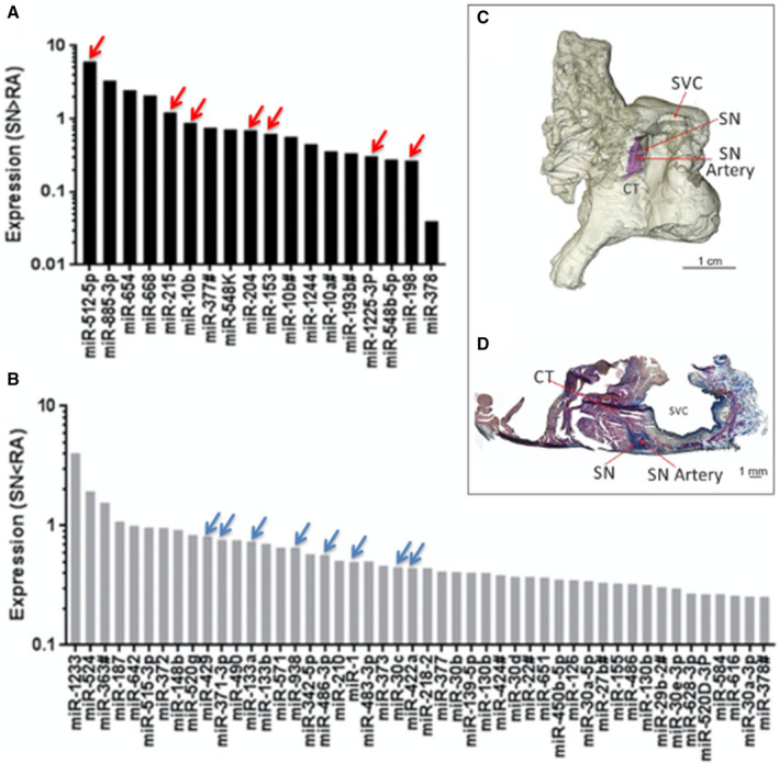
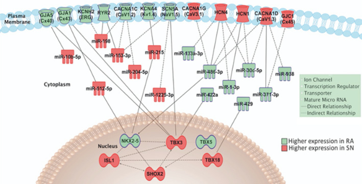
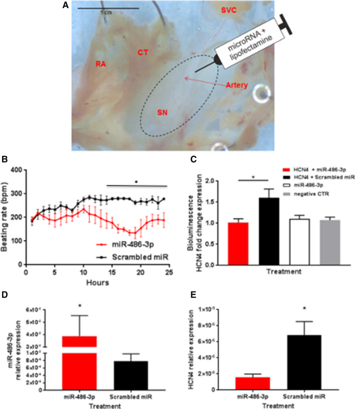
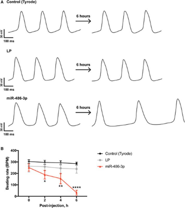
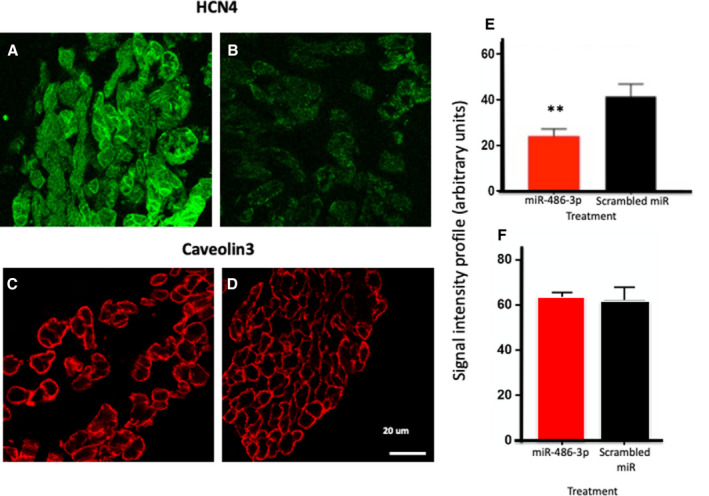
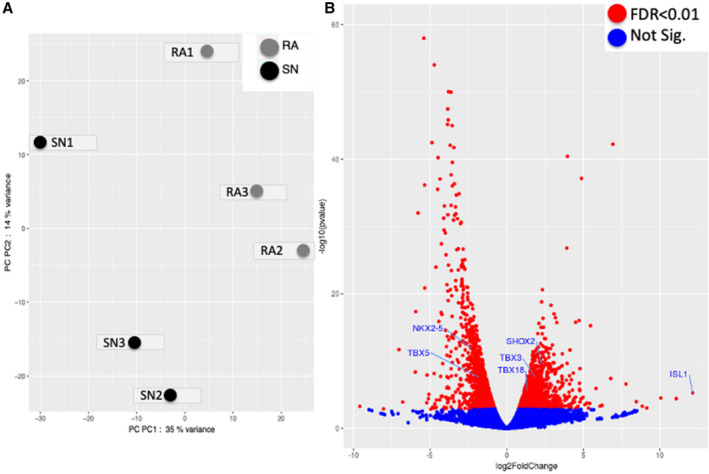
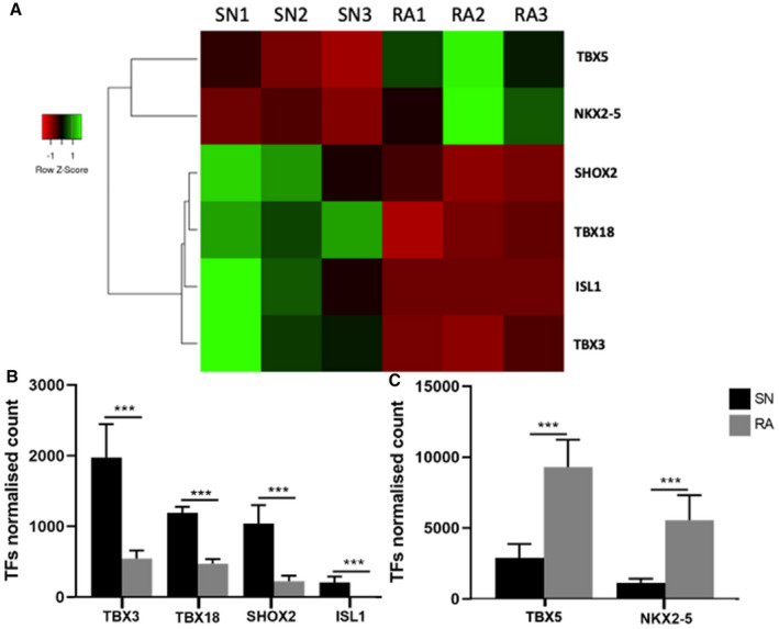
Background The sinus node (SN) is the primary pacemaker of the heart. SN myocytes possess distinctive action potential morphology with spontaneous diastolic depolarization because of a unique expression of ion channels and Ca-handling proteins. MicroRNAs (miRs) inhibit gene expression. The role of miRs in controlling the expression of genes responsible for human SN pacemaking and conduction has not been explored. The aim of this study was to determine miR expression profile of the human SN as compared with that of non-pacemaker atrial muscle. Methods and Results SN and atrial muscle biopsies were obtained from donor or post-mortem hearts (n=10), histology/immunolabeling were used to characterize the tissues, TaqMan Human MicroRNA Arrays were used to measure 754 miRs, Ingenuity Pathway Analysis was used to identify miRs controlling SN pacemaker gene expression. Eighteen miRs were significantly more and 48 significantly less abundant in the SN than atrial muscle. The most interesting miR was miR-486-3p predicted to inhibit expression of pacemaking channels: HCN1 (hyperpolarization-activated cyclic nucleotide-gated 1), HCN4, voltage-gated calcium channel (Ca)1.3, and Ca3.1. A luciferase reporter gene assay confirmed that miR-486-3p can control HCN4 expression via its 3' untranslated region. In SN preparations, transfection with miR-486-3p reduced the beating rate by ≈35±5% (<0.05) and HCN4 expression (<0.05). Conclusions The human SN possesses a unique pattern of expression of miRs predicted to target functionally important genes. miR-486-3p has an important role in SN pacemaker activity by targeting HCN4, making it a potential target for therapeutic treatment of SN disease such as sinus tachycardia.
窦房结(SN)是心脏的主要起搏点。由于离子通道和钙处理蛋白的独特表达,SN 心肌细胞具有独特的动作电位形态和自发性舒张去极化。MicroRNAs(miRs)抑制基因表达。miRs 在控制负责人类 SN 起搏和传导的基因表达方面的作用尚未得到探索。本研究旨在确定与非起搏心房肌相比,人 SN 的 miR 表达谱。
从供体或死后心脏(n=10)获得 SN 和心房肌活检,组织学/免疫标记用于对组织进行特征描述,TaqMan 人类 MicroRNA 阵列用于测量 754 个 miR,Ingenuity 通路分析用于识别控制 SN 起搏基因表达的 miR。18 个 miR 在 SN 中的表达明显高于心房肌,而 48 个 miR 的表达明显低于心房肌。最有趣的 miR 是 miR-486-3p,它被预测可以抑制起搏通道的表达:HCN1(超极化激活环核苷酸门控 1)、HCN4、电压门控钙通道(Ca)1.3 和 Ca3.1。荧光素酶报告基因检测证实 miR-486-3p 可以通过其 3'非翻译区控制 HCN4 的表达。在 SN 制剂中,miR-486-3p 的转染使搏动率降低约 35±5%(<0.05)和 HCN4 表达(<0.05)。
人类 SN 具有独特的 miR 表达模式,这些 miR 预测靶向功能重要的基因。miR-486-3p 通过靶向 HCN4 在 SN 起搏活动中具有重要作用,使其成为治疗 SN 疾病(如窦性心动过速)的潜在治疗靶点。