Department of Cardiology, Pulmonology and Vascular Medicine, Heinrich Heine University Düsseldorf, Moorenstraße 5, Düsseldorf, 40225, Germany.
Philips Healthcare, Hamburg, Germany.
Eur Radiol. 2021 May;31(5):2768-2777. doi: 10.1007/s00330-020-07289-4. Epub 2020 Oct 15.
Distinguishing hypertrophic cardiomyopathy (HCM) from left ventricular hypertrophy (LVH) due to systematic training (athlete's heart, AH) from morphologic assessment remains challenging. The purpose of this study was to examine the role of T2 mapping and deformation imaging obtained by cardiovascular magnetic resonance (CMR) to discriminate AH from HCM with (HOCM) or without outflow tract obstruction (HNCM).
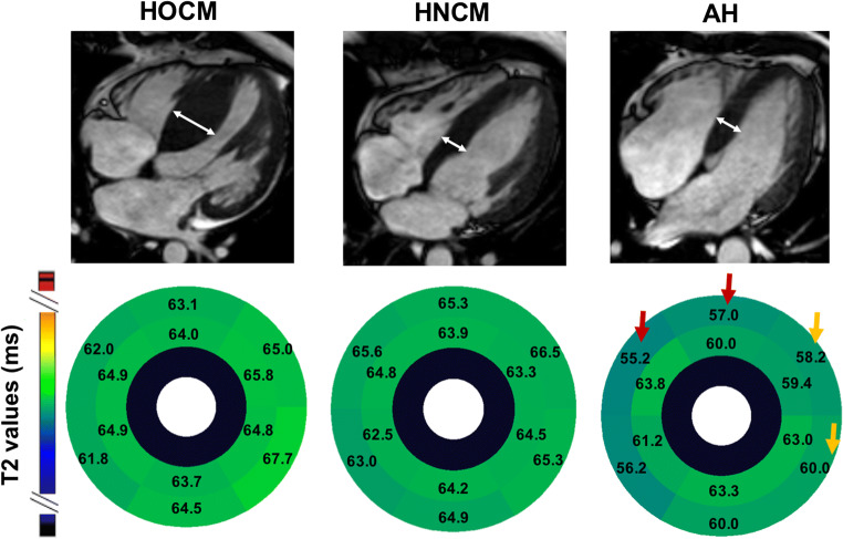
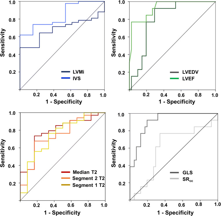
Thirty-three patients with HOCM, 9 with HNCM, 13 strength-trained athletes as well as individual age- and gender-matched controls received CMR. For T2 mapping, GRASE-derived multi-echo images were obtained and analyzed using dedicated software. Besides T2 mapping analyses, left ventricular (LV) dimensional and functional parameters were obtained including LV mass per body surface area (LVMi), interventricular septum thickness (IVS), and global longitudinal strain (GLS).
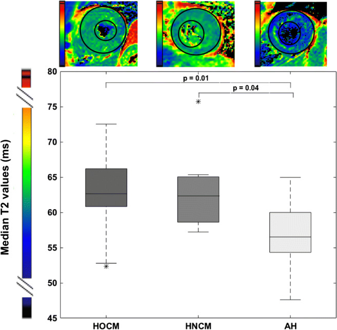
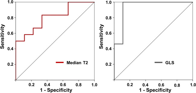
While LVMi was not significantly different, IVS was thickened in HOCM patients compared to athlete's. Absolute values of GLS were significantly increased in patients with HOCM/HNCM compared to AH. Median T2 values were elevated compared to controls except in athlete's heart. ROC analysis revealed T2 values (AUC 0.78) and GLS (AUC 0.91) as good parameters to discriminate AH from overall HNCM/HOCM.
Discrimination of pathologic from non-pathologic LVH has implications for risk assessment of competitive sports in athletes. Multiparametric CMR with parametric T2 mapping and deformation imaging may add information to distinguish AH from LVH due to HCM.
• Structural analyses using T2 mapping cardiovascular magnetic resonance imaging (CMR) may help to further distinguish myocardial diseases. • To differentiate pathologic from non-pathologic left ventricular hypertrophy, CMR including T2 mapping was obtained in patients with hypertrophic obstructive/non-obstructive cardiomyopathy (HOCM/HNCM) as well as in strength-trained athletes. • Elevated median T2 values in HOCM/HNCM compared with athlete's may add information to distinguish athlete's heart from pathologic left ventricular hypertrophy.
从形态学评估上区分由于系统训练(运动员心脏,AH)引起的左心室肥厚(LVH)与肥厚型心肌病(HCM)仍然具有挑战性。本研究旨在通过心血管磁共振(CMR)检查 T2 映射和变形成像的作用,来区分伴有(HOCM)或不伴有流出道梗阻(HNCM)的 HCM 与 AH。
33 例 HOCM 患者、9 例 HNCM 患者、13 名力量训练运动员以及年龄和性别匹配的个体对照组接受 CMR 检查。使用 GRASE 衍生的多回波图像进行 T2 映射,并使用专用软件进行分析。除 T2 映射分析外,还获得了左心室(LV)尺寸和功能参数,包括 LV 质量/体表面积(LVMi)、室间隔厚度(IVS)和整体纵向应变(GLS)。
虽然 LVMi 没有显著差异,但 HOCM 患者的 IVS 比运动员的更厚。与 AH 相比,HOCM/HNCM 患者的 GLS 绝对值明显增加。与对照组相比,除了运动员心脏外,HOCM/HNCM 患者的 T2 值中位数升高。ROC 分析显示 T2 值(AUC 0.78)和 GLS(AUC 0.91)是区分 AH 与整体 HNCM/HOCM 的良好参数。
病理性与非病理性 LVH 的区分对运动员竞技运动的风险评估具有重要意义。使用 T2 映射心血管磁共振成像(CMR)的多参数 CMR 可能有助于进一步区分心肌疾病。
使用 T2 映射心血管磁共振成像(CMR)的结构分析可能有助于进一步区分心肌疾病。
为了区分病理性与非病理性左心室肥厚,对患有肥厚型梗阻性/非梗阻性心肌病(HOCM/HNCM)的患者以及力量训练运动员进行了包括 T2 映射的 CMR 检查。
与运动员心脏相比,HOCM/HNCM 中的中位数 T2 值升高可能有助于将运动员心脏与病理性左心室肥厚区分开来。