• 文献检索
  • 文档翻译
  • 深度研究
  • 学术资讯
  • Suppr Zotero 插件Zotero 插件
  • 邀请有礼
  • 套餐&价格
  • 历史记录
应用&插件
Suppr Zotero 插件Zotero 插件浏览器插件Mac 客户端Windows 客户端微信小程序
定价
高级版会员购买积分包购买API积分包
服务
文献检索文档翻译深度研究API 文档MCP 服务
关于我们
关于 Suppr公司介绍联系我们用户协议隐私条款
关注我们

Suppr 超能文献

核心技术专利:CN118964589B侵权必究
粤ICP备2023148730 号-1Suppr @ 2026

文献检索

告别复杂PubMed语法,用中文像聊天一样搜索,搜遍4000万医学文献。AI智能推荐,让科研检索更轻松。

立即免费搜索

文件翻译

保留排版,准确专业,支持PDF/Word/PPT等文件格式,支持 12+语言互译。

免费翻译文档

深度研究

AI帮你快速写综述,25分钟生成高质量综述,智能提取关键信息,辅助科研写作。

立即免费体验

HARDI-ZOOMit 方案提高了无症状性脊髓病微观结构变化的特异性。

HARDI-ZOOMit protocol improves specificity to microstructural changes in presymptomatic myelopathy.

机构信息

Department of Biomedical Engineering, University Hospital Olomouc, 779 00, Olomouc, Czech Republic.

Department of Neurology, Palacký University, 779 00, Olomouc, Czech Republic.

出版信息

Sci Rep. 2020 Oct 16;10(1):17529. doi: 10.1038/s41598-020-70297-3.

DOI:10.1038/s41598-020-70297-3
PMID:33067520
原文链接:https://pmc.ncbi.nlm.nih.gov/articles/PMC7567840/
Abstract

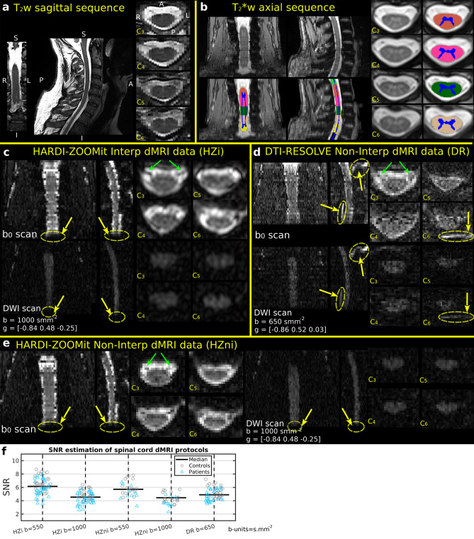
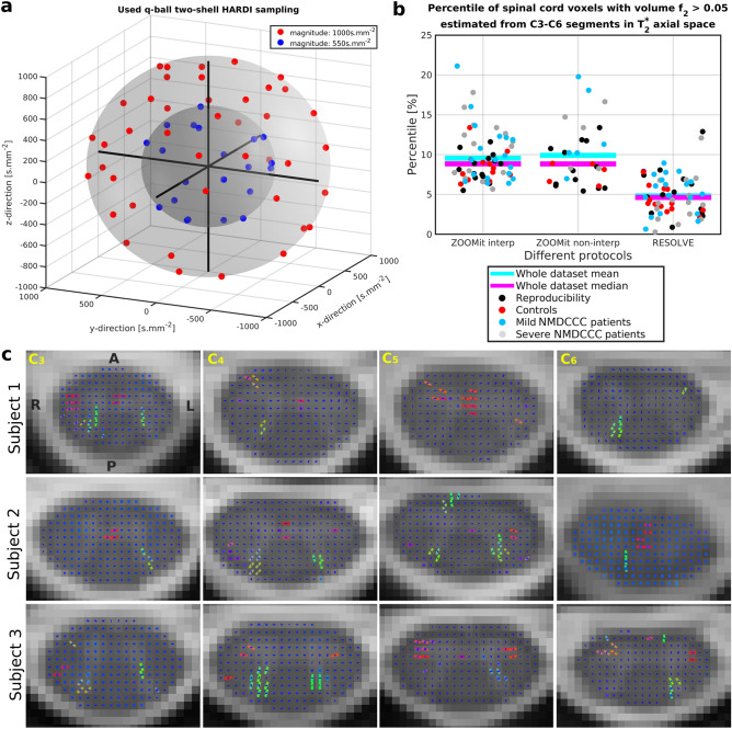
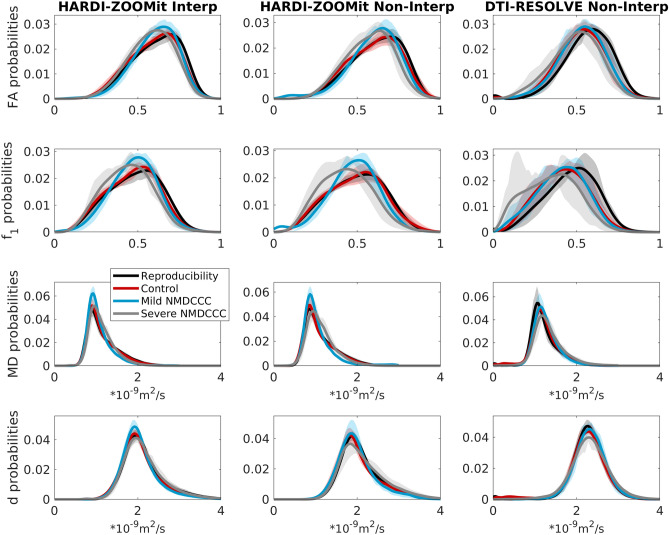
Diffusion magnetic resonance imaging (dMRI) proved promising in patients with non-myelopathic degenerative cervical cord compression (NMDCCC), i.e., without clinically manifested myelopathy. Aim of the study is to present a fast multi-shell HARDI-ZOOMit dMRI protocol and validate its usability to detect microstructural myelopathy in NMDCCC patients. In 7 young healthy volunteers, 13 age-comparable healthy controls, 18 patients with mild NMDCCC and 15 patients with severe NMDCCC, the protocol provided higher signal-to-noise ratio, enhanced visualization of white/gray matter structures in microstructural maps, improved dMRI metric reproducibility, preserved sensitivity (SE = 87.88%) and increased specificity (SP = 92.31%) of control-patient group differences when compared to DTI-RESOLVE protocol (SE = 87.88%, SP = 76.92%). Of the 56 tested microstructural parameters, HARDI-ZOOMit yielded significant patient-control differences in 19 parameters, whereas in DTI-RESOLVE data, differences were observed in 10 parameters, with mostly lower robustness. Novel marker the white-gray matter diffusivity gradient demonstrated the highest separation. HARDI-ZOOMit protocol detected larger number of crossing fibers (5-15% of voxels) with physiologically plausible orientations than DTI-RESOLVE protocol (0-8% of voxels). Crossings were detected in areas of dorsal horns and anterior white commissure. HARDI-ZOOMit protocol proved to be a sensitive and practical tool for clinical quantitative spinal cord imaging.

摘要

弥散磁共振成像(dMRI)在非脊髓型退行性颈椎脊髓压迫症(NMDCCC)患者中显示出良好的应用前景,即无临床表现的脊髓病。本研究旨在提出一种快速多壳 HARDI-ZOOMit dMRI 方案,并验证其在检测 NMDCCC 患者微结构脊髓病中的可用性。在 7 名年轻健康志愿者、13 名年龄匹配的健康对照者、18 名轻度 NMDCCC 患者和 15 名重度 NMDCCC 患者中,该方案提供了更高的信噪比,增强了微结构图谱中白/灰质结构的可视化效果,提高了 dMRI 指标的可重复性,保持了对患者和对照组差异的敏感性(SE = 87.88%),并提高了特异性(SP = 92.31%),与 DTI-RESOLVE 方案(SE = 87.88%,SP = 76.92%)相比。在 56 个测试的微结构参数中,HARDI-ZOOMit 在 19 个参数中产生了显著的患者-对照组差异,而在 DTI-RESOLVE 数据中,仅在 10 个参数中观察到差异,且其稳健性较低。新的标志物白质-灰质扩散梯度显示出最高的分离度。HARDI-ZOOMit 方案检测到的交叉纤维(5-15%的体素)数量多于 DTI-RESOLVE 方案(0-8%的体素),且具有更合理的生理方向。这些交叉纤维在背角和前白质联合区被检测到。HARDI-ZOOMit 方案被证明是一种敏感而实用的临床定量脊髓成像工具。

https://cdn.ncbi.nlm.nih.gov/pmc/blobs/c00b/7567840/f9b80054d487/41598_2020_70297_Fig4_HTML.jpg
https://cdn.ncbi.nlm.nih.gov/pmc/blobs/c00b/7567840/4598206eea1e/41598_2020_70297_Fig1_HTML.jpg
https://cdn.ncbi.nlm.nih.gov/pmc/blobs/c00b/7567840/e98e01913f14/41598_2020_70297_Fig3_HTML.jpg
https://cdn.ncbi.nlm.nih.gov/pmc/blobs/c00b/7567840/f9b80054d487/41598_2020_70297_Fig4_HTML.jpg
https://cdn.ncbi.nlm.nih.gov/pmc/blobs/c00b/7567840/4598206eea1e/41598_2020_70297_Fig1_HTML.jpg
https://cdn.ncbi.nlm.nih.gov/pmc/blobs/c00b/7567840/e98e01913f14/41598_2020_70297_Fig3_HTML.jpg
https://cdn.ncbi.nlm.nih.gov/pmc/blobs/c00b/7567840/f9b80054d487/41598_2020_70297_Fig4_HTML.jpg

相似文献

1
HARDI-ZOOMit protocol improves specificity to microstructural changes in presymptomatic myelopathy.HARDI-ZOOMit 方案提高了无症状性脊髓病微观结构变化的特异性。
Sci Rep. 2020 Oct 16;10(1):17529. doi: 10.1038/s41598-020-70297-3.
2
Diffusion tensor imaging of somatosensory tract in cervical spondylotic myelopathy and its link with electrophysiological evaluation.脊髓型颈椎病体感通路的扩散张量成像及其与电生理评估的关联
Spine J. 2014 Aug 1;14(8):1493-500. doi: 10.1016/j.spinee.2013.08.052. Epub 2013 Oct 4.
3
Neurodegeneration in the Spinal Ventral Horn Prior to Motor Impairment in Cervical Spondylotic Myelopathy.脊髓型颈椎病运动功能障碍之前脊髓腹角的神经变性
J Neurotrauma. 2017 Aug 1;34(15):2329-2334. doi: 10.1089/neu.2017.4980. Epub 2017 Jun 27.
4
Accuracy of Diffusion Tensor Imaging for Diagnosing Cervical Spondylotic Myelopathy in Patients Showing Spinal Cord Compression.扩散张量成像对显示脊髓受压的颈椎病性脊髓病患者的诊断准确性
Korean J Radiol. 2015 Nov-Dec;16(6):1303-12. doi: 10.3348/kjr.2015.16.6.1303. Epub 2015 Oct 26.
5
Tracking White and Gray Matter Degeneration along the Spinal Cord Axis in Degenerative Cervical Myelopathy.追踪退行性颈椎脊髓病中脊髓轴上的白质和灰质退变。
J Neurotrauma. 2021 Nov 1;38(21):2978-2987. doi: 10.1089/neu.2021.0148. Epub 2021 Aug 3.
6
Orientation entropy analysis of diffusion tensor in healthy and myelopathic spinal cord.健康与脊髓病变脊髓扩散张量各向异性熵分析。
Neuroimage. 2011 Oct 15;58(4):1028-33. doi: 10.1016/j.neuroimage.2011.06.072. Epub 2011 Jul 18.
7
Predictors of symptomatic myelopathy in degenerative cervical spinal cord compression.退变性颈椎脊髓压迫症中症状性脊髓病的预测因素。
Brain Behav. 2017 Aug 11;7(9):e00797. doi: 10.1002/brb3.797. eCollection 2017 Sep.
8
Reproducibility, temporal stability, and functional correlation of diffusion MR measurements within the spinal cord in patients with asymptomatic cervical stenosis or cervical myelopathy.无症状性颈椎管狭窄或脊髓型颈椎病患者脊髓内扩散磁共振测量的可重复性、时间稳定性及功能相关性
J Neurosurg Spine. 2018 May;28(5):472-480. doi: 10.3171/2017.7.SPINE176. Epub 2018 Feb 9.
9
Microstructural Changes in Compressed Cervical Spinal Cord Are Consistent With Clinical Symptoms and Symptom Duration.受压颈椎脊髓的微观结构变化与临床症状和症状持续时间一致。
Spine (Phila Pa 1976). 2020 Aug 15;45(16):E999-E1005. doi: 10.1097/BRS.0000000000003480.
10
Correlation of magnetic resonance diffusion tensor imaging and clinical findings of cervical myelopathy.磁共振弥散张量成像与颈椎病临床相关性研究。
Spine J. 2013 Aug;13(8):867-76. doi: 10.1016/j.spinee.2013.02.005. Epub 2013 Mar 21.

引用本文的文献

1
Body size and intracranial volume interact with the structure of the central nervous system: A multi-center in vivo neuroimaging study.身体大小与颅内体积与中枢神经系统结构相互作用:一项多中心活体神经影像学研究。
Imaging Neurosci (Camb). 2025 May 7;3. doi: 10.1162/imag_a_00559. eCollection 2025.
2
A Systematic Review of Current Terminology for Conditions Preceding Degenerative Cervical Myelopathy: Evidence Synthesis to Inform an AO Spine Expert Opinion Statement.退行性颈椎脊髓病前驱病症当前术语的系统评价:为AO脊柱专家意见声明提供信息的证据综合
Global Spine J. 2025 Apr 30:21925682251339480. doi: 10.1177/21925682251339480.
3

本文引用的文献

1
Tract-specific analysis improves sensitivity of spinal cord diffusion MRI to cross-sectional and longitudinal changes in amyotrophic lateral sclerosis.特定区域分析提高了脊髓扩散磁共振成像对肌萎缩侧索硬化症横断面和纵向变化的敏感性。
Commun Biol. 2020 Jul 10;3(1):370. doi: 10.1038/s42003-020-1093-z.
2
Longitudinal diffusion MRI as surrogate outcome measure for myelopathy in adrenoleukodystrophy.纵向弥散 MRI 作为肾上腺脑白质营养不良性脊髓病的替代结局测量指标。
Neurology. 2019 Dec 3;93(23):e2133-e2143. doi: 10.1212/WNL.0000000000008572. Epub 2019 Nov 12.
3
Diffusion MRI microstructural models in the cervical spinal cord - Application, normative values, and correlations with histological analysis.
Evidence-based commentary on the diagnosis, management, and further research of degenerative cervical spinal cord compression in the absence of clinical symptoms of myelopathy.
关于无脊髓病临床症状的退行性颈椎脊髓压迫症的诊断、管理及进一步研究的循证评论
Front Neurol. 2024 May 10;15:1341371. doi: 10.3389/fneur.2024.1341371. eCollection 2024.
4
Body size interacts with the structure of the central nervous system: A multi-center in vivo neuroimaging study.身体大小与中枢神经系统结构相互作用:一项多中心活体神经影像学研究。
bioRxiv. 2024 May 1:2024.04.29.591421. doi: 10.1101/2024.04.29.591421.
5
Reproducible Spinal Cord Quantitative MRI Analysis with the Spinal Cord Toolbox.可重现的脊髓定量 MRI 分析——脊髓工具箱。
Magn Reson Med Sci. 2024 Jul 1;23(3):307-315. doi: 10.2463/mrms.rev.2023-0159. Epub 2024 Mar 12.
6
Evaluations of the diagnostic performance of ZOOMit diffusion-weighted imaging and conventional diffusion-weighted imaging for breast lesions.ZOOMit扩散加权成像与传统扩散加权成像对乳腺病变诊断性能的评估。
Quant Imaging Med Surg. 2023 Dec 1;13(12):8478-8488. doi: 10.21037/qims-23-415. Epub 2023 Nov 7.
7
Spinal cord magnetic resonance imaging and spectroscopy detect early-stage alterations and disease progression in Friedreich ataxia.脊髓磁共振成像和波谱分析可检测弗里德赖希共济失调的早期改变和疾病进展。
Brain Commun. 2022 Oct 3;4(5):fcac246. doi: 10.1093/braincomms/fcac246. eCollection 2022.
8
Quantitative MR Markers in Non-Myelopathic Spinal Cord Compression: A Narrative Review.非脊髓型脊髓压迫症的定量磁共振成像标志物:一篇叙述性综述
J Clin Med. 2022 Apr 20;11(9):2301. doi: 10.3390/jcm11092301.
9
Semi-automated detection of cervical spinal cord compression with the Spinal Cord Toolbox.使用脊髓工具箱半自动检测颈脊髓压迫症
Quant Imaging Med Surg. 2022 Apr;12(4):2261-2279. doi: 10.21037/qims-21-782.
10
Comparative Study between ZOOMit and Conventional Intravoxel Incoherent Motion MRI for Assessing Parotid Gland Abnormalities in Patients with Early- or Mid-Stage Sjögren's Syndrome.ZOOMit 与常规体素内不相干运动 MRI 评估早中期干燥综合征患者腮腺异常的对比研究。
Korean J Radiol. 2022 Apr;23(4):455-465. doi: 10.3348/kjr.2021.0695. Epub 2022 Mar 8.
弥散磁共振成像在颈脊髓微观结构模型中的应用——与组织学分析的相关性及正常值。
Neuroimage. 2019 Nov 1;201:116026. doi: 10.1016/j.neuroimage.2019.116026. Epub 2019 Jul 19.
4
Automatic segmentation of the spinal cord and intramedullary multiple sclerosis lesions with convolutional neural networks.基于卷积神经网络的脊髓和髓内多发性硬化病变自动分割。
Neuroimage. 2019 Jan 1;184:901-915. doi: 10.1016/j.neuroimage.2018.09.081. Epub 2018 Oct 6.
5
Postmortem diffusion MRI of the entire human spinal cord at microscopic resolution.高分辨率全脊髓弥散磁共振成像的尸检研究
Neuroimage Clin. 2018 Mar 27;18:963-971. doi: 10.1016/j.nicl.2018.03.029. eCollection 2018.
6
Monitoring for myelopathic progression with multiparametric quantitative MRI.多参数定量 MRI 监测脊髓病进展。
PLoS One. 2018 Apr 17;13(4):e0195733. doi: 10.1371/journal.pone.0195733. eCollection 2018.
7
Spinal cord gray matter segmentation using deep dilated convolutions.使用深度扩张卷积进行脊髓灰质分割。
Sci Rep. 2018 Apr 13;8(1):5966. doi: 10.1038/s41598-018-24304-3.
8
Can microstructural MRI detect subclinical tissue injury in subjects with asymptomatic cervical spinal cord compression? A prospective cohort study.微观结构磁共振成像能否检测无症状性颈脊髓压迫患者的亚临床组织损伤?一项前瞻性队列研究。
BMJ Open. 2018 Apr 13;8(4):e019809. doi: 10.1136/bmjopen-2017-019809.
9
Reproducibility, temporal stability, and functional correlation of diffusion MR measurements within the spinal cord in patients with asymptomatic cervical stenosis or cervical myelopathy.无症状性颈椎管狭窄或脊髓型颈椎病患者脊髓内扩散磁共振测量的可重复性、时间稳定性及功能相关性
J Neurosurg Spine. 2018 May;28(5):472-480. doi: 10.3171/2017.7.SPINE176. Epub 2018 Feb 9.
10
Feasibility of single-shot multi-level multi-angle diffusion tensor imaging of the human cervical spinal cord at 7T.7T 下人体颈脊髓单次多层多角度扩散张量成像的可行性
Magn Reson Med. 2018 Sep;80(3):947-957. doi: 10.1002/mrm.27087. Epub 2018 Jan 25.