Lei Yiming, Fu Xuekun, Li Pengyu, Lin Sixiong, Yan Qinnan, Lai Yumei, Liu Xin, Wang Yishu, Bai Xiaochun, Liu Chuanju, Chen Di, Zou Xuenong, Cao Xu, Cao Huiling, Xiao Guozhi
Guangdong Provincial Key Laboratory of Cell Microenvironment and Disease Research, Shenzhen Key Laboratory of Cell Microenvironment, and School of Medicine, Southern University of Science and Technology, Shenzhen, 518055 China.
Department of Spine Surgery, Orthopedic Research Institute, The First Affiliated Hospital of Sun Yat-sen University, Guangdong Provincial Key Laboratory of Orthopedics and Traumatology, Guangzhou, 510080 China.
Bone Res. 2020 Oct 13;8:37. doi: 10.1038/s41413-020-00108-y. eCollection 2020.
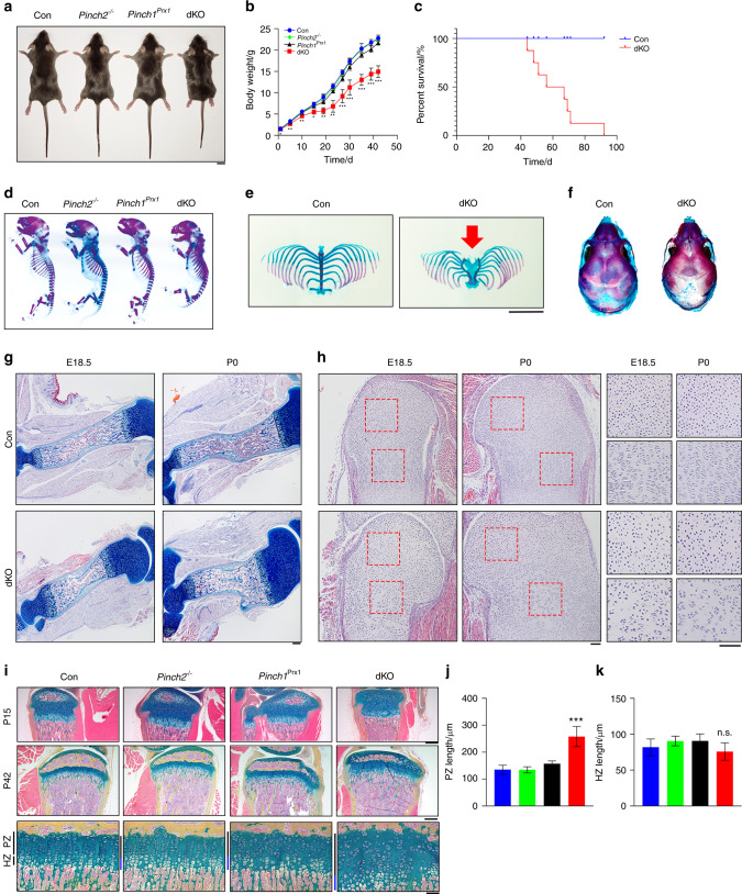
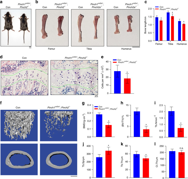
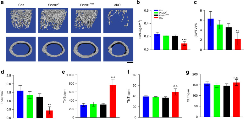
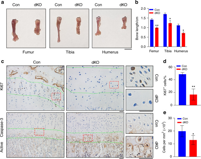
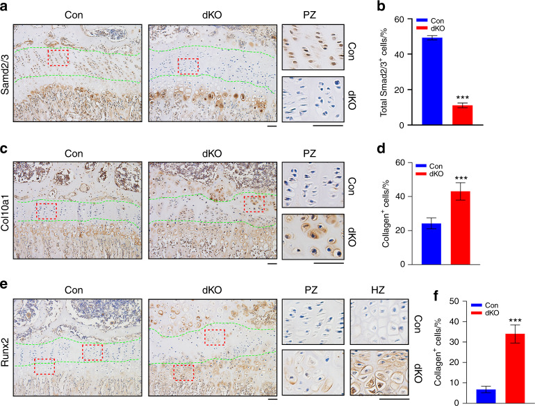
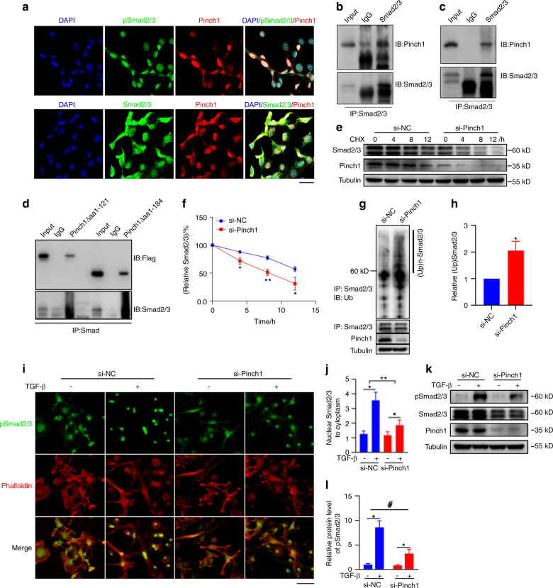
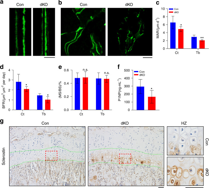
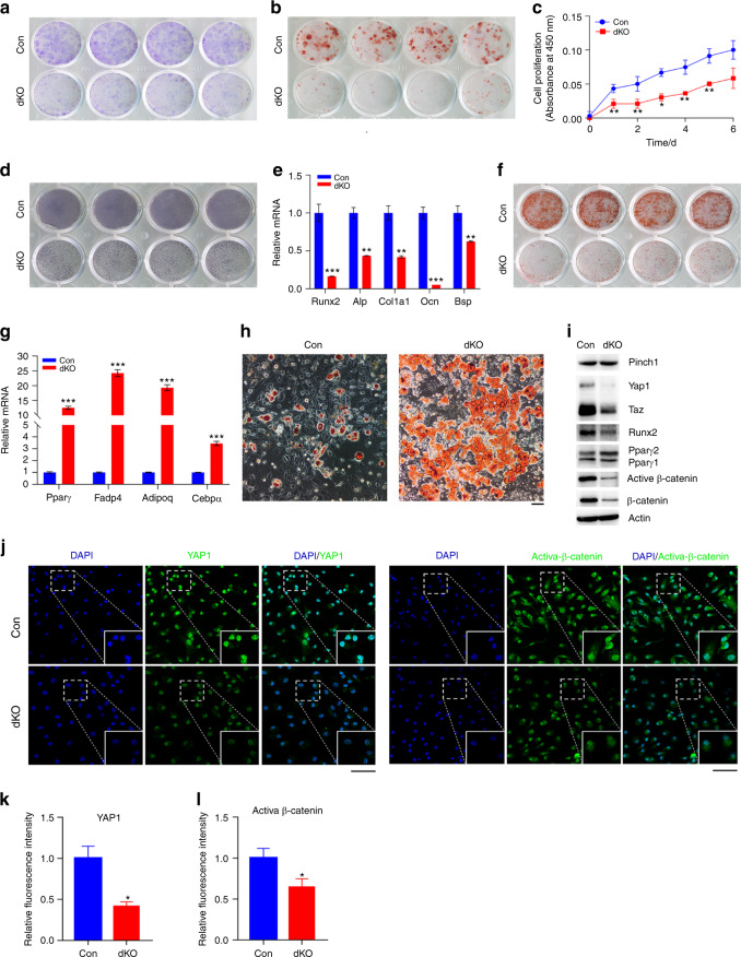
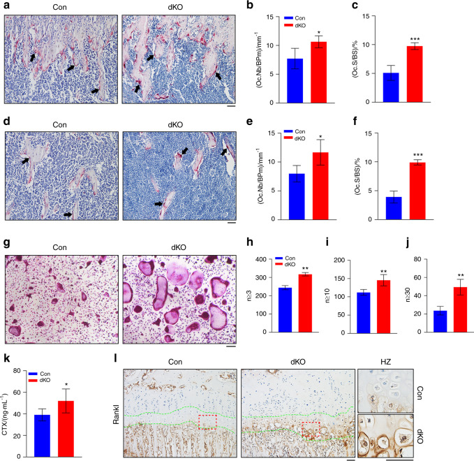
The LIM domain-containing proteins Pinch1/2 regulate integrin activation and cell-extracellular matrix interaction and adhesion. Here, we report that deleting Pinch1 in limb mesenchymal stem cells (MSCs) and Pinch2 globally (double knockout; dKO) in mice causes severe chondrodysplasia, while single mutant mice do not display marked defects. Pinch deletion decreases chondrocyte proliferation, accelerates cell differentiation and disrupts column formation. Pinch loss drastically reduces Smad2/3 protein expression in proliferative zone (PZ) chondrocytes and increases Runx2 and Col10a1 expression in both PZ and hypertrophic zone (HZ) chondrocytes. Pinch loss increases sclerostin and Rankl expression in HZ chondrocytes, reduces bone formation, and increases bone resorption, leading to low bone mass. In vitro studies revealed that Pinch1 and Smad2/3 colocalize in the nuclei of chondrocytes. Through its C-terminal region, Pinch1 interacts with Smad2/3 proteins. Pinch loss increases Smad2/3 ubiquitination and degradation in primary bone marrow stromal cells (BMSCs). Pinch loss reduces TGF-β-induced Smad2/3 phosphorylation and nuclear localization in primary BMSCs. Interestingly, compared to those from single mutant mice, BMSCs from dKO mice express dramatically lower protein levels of β-catenin and Yap1/Taz and display reduced osteogenic but increased adipogenic differentiation capacity. Finally, ablating Pinch1 in chondrocytes and Pinch2 globally causes severe osteopenia with subtle limb shortening. Collectively, our findings demonstrate critical roles for Pinch1/2 and a functional redundancy of both factors in the control of chondrogenesis and bone mass through distinct mechanisms.
含LIM结构域的蛋白Pinch1/2调节整合素激活以及细胞与细胞外基质的相互作用和黏附。在此,我们报告,在小鼠的肢体间充质干细胞(MSC)中删除Pinch1并全局删除Pinch2(双敲除;dKO)会导致严重的软骨发育不全,而单突变小鼠未表现出明显缺陷。Pinch缺失会降低软骨细胞增殖,加速细胞分化并破坏柱状结构形成。Pinch缺失会大幅降低增殖区(PZ)软骨细胞中Smad2/3蛋白表达,并增加PZ和肥大区(HZ)软骨细胞中Runx2和Col10a1的表达。Pinch缺失会增加HZ软骨细胞中硬化蛋白和Rankl表达,减少骨形成,并增加骨吸收,导致骨量降低。体外研究表明,Pinch1和Smad2/3共定位于软骨细胞核中。Pinch1通过其C末端区域与Smad2/3蛋白相互作用。Pinch缺失会增加原代骨髓基质细胞(BMSC)中Smad2/3的泛素化和降解。Pinch缺失会降低原代BMSC中TGF-β诱导的Smad2/3磷酸化和核定位。有趣的是,与单突变小鼠的BMSC相比,dKO小鼠的BMSC表达的β-连环蛋白和Yap1/Taz蛋白水平显著降低,并且成骨分化能力降低但脂肪生成分化能力增加。最后,在软骨细胞中消融Pinch1并全局消融Pinch2会导致严重的骨质减少并伴有轻微的肢体缩短。总的来说,我们的研究结果证明了Pinch1/2的关键作用以及这两种因子在通过不同机制控制软骨生成和骨量方面的功能冗余。