Xue Yujia, Davison Ian G, Boas David A, Tian Lei
Department of Electrical and Computer Engineering, Boston University, MA 02215, USA.
Department of Biology, Boston University, MA 02215, USA.
Sci Adv. 2020 Oct 21;6(43). doi: 10.1126/sciadv.abb7508. Print 2020 Oct.
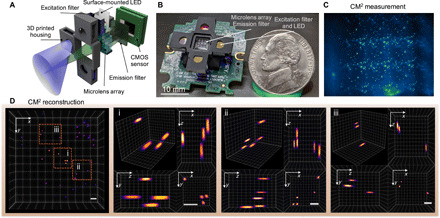
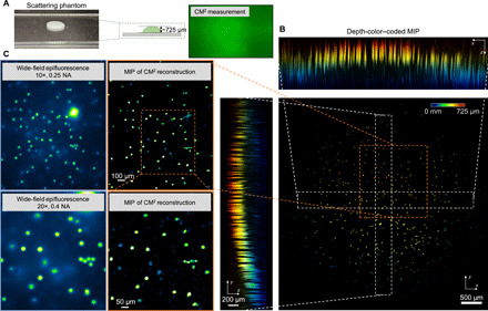
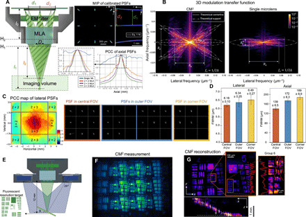
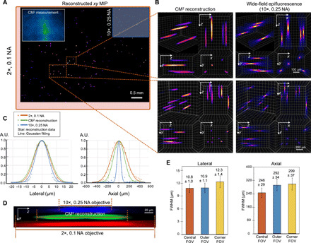
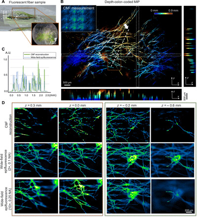
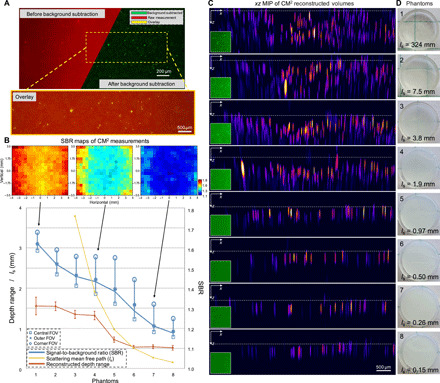
Fluorescence microscopes are indispensable to biology and neuroscience. The need for recording in freely behaving animals has further driven the development in miniaturized microscopes (miniscopes). However, conventional microscopes/miniscopes are inherently constrained by their limited space-bandwidth product, shallow depth of field (DOF), and inability to resolve three-dimensional (3D) distributed emitters. Here, we present a Computational Miniature Mesoscope (CM) that overcomes these bottlenecks and enables single-shot 3D imaging across an 8 mm by 7 mm field of view and 2.5-mm DOF, achieving 7-μm lateral resolution and better than 200-μm axial resolution. The CM features a compact lightweight design that integrates a microlens array for imaging and a light-emitting diode array for excitation. Its expanded imaging capability is enabled by computational imaging that augments the optics by algorithms. We experimentally validate the mesoscopic imaging capability on 3D fluorescent samples. We further quantify the effects of scattering and background fluorescence on phantom experiments.
荧光显微镜对于生物学和神经科学来说不可或缺。在自由活动的动物中进行记录的需求进一步推动了小型显微镜(微型显微镜)的发展。然而,传统显微镜/微型显微镜固有地受到其有限的空间带宽积、浅景深(DOF)以及无法分辨三维(3D)分布的发光体的限制。在此,我们展示了一种计算微型介观显微镜(CM),它克服了这些瓶颈,能够在8毫米×7毫米的视场和2.5毫米的景深范围内进行单次3D成像,实现7微米的横向分辨率和优于200微米的轴向分辨率。CM具有紧凑轻便的设计,集成了用于成像的微透镜阵列和用于激发的发光二极管阵列。其扩展的成像能力是通过计算成像实现的,该成像通过算法增强了光学性能。我们通过实验验证了对3D荧光样本的介观成像能力。我们还在模型实验中进一步量化了散射和背景荧光的影响。