From the Department of Neurology (M.P., M.M.E.M., M.M., S.C., K.P., M.I.), Icahn School of Medicine at Mount Sinai, NY; Aix-Marseille Univ (M.M.E.M.), CNRS, CRMBM; APHM (M.M.E.M.), Hôpital de la Timone, CEMEREM, Marseille, France; Department of Informatics (M.M.), Bioengineering, Robotics and Systems Engineering (DIBRIS) and Machine Learning Genoa Center (MaLGa), University of Genoa; Department of Advanced Biomedical Sciences (S.C.), University of Naples "Federico II", Italy; Department of Radiology (L.F.), Icahn School of Medicine at Mount Sinai, NY; Department of Neurosciences, Rehabilitation, Ophthalmology, Genetics (M.I.), Maternal and Child Health (DINOGMI) and Center of Excellence for Biomedical Research, University of Genoa; and Ospedale Policlinico San Martino-IRCCS (M.I.), Genoa, Italy.
Neurol Neuroimmunol Neuroinflamm. 2020 Oct 21;7(6). doi: 10.1212/NXI.0000000000000900. Print 2020 Nov.
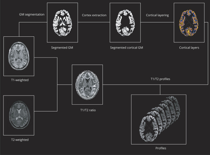
In this observational study, we explored cortical structure as function of cortical depth through a laminar analysis of the T1/T2-weighted (T1w/T2w) ratio, which has been related to dendrite density in ex vivo brain tissue specimens of patients with MS.
In 39 patients (22 relapsing-remitting, 13 female, age 41.1 ± 10.6 years; 17 progressive, 11 female, age 54.1 ± 9.9 years) and 21 healthy controls (8 female, , age 41.6 ± 10.6 years), we performed a voxel-wise analysis of T1w/T2w ratio maps from high-resolution 7T images from the subpial surface to the gray matter/white matter boundary. Six layers were sampled to ensure accuracy based on mean cortical thickness and image resolution.
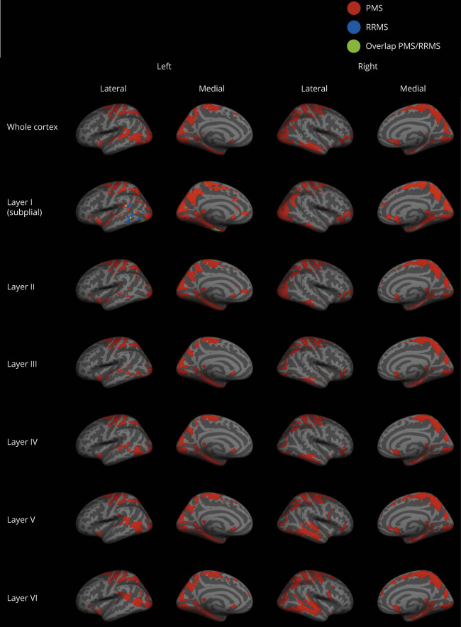
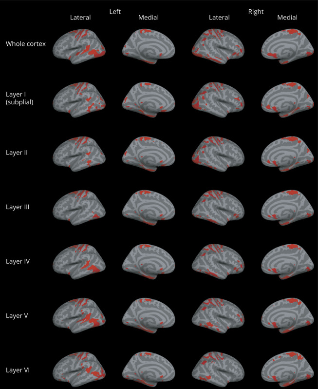
At the voxel-wise comparison ( < 0.05, family wise error rate corrected), the whole MS group showed lower T1w/T2w ratio values than controls, both when considering the entire cortex and each individual layer, with peaks occurring in the fusiform, temporo-occipital, and superior and middle frontal cortex. In relapsing-remitting patients, differences in the T1w/T2w ratio were only identified in the subpial layer, with the peak occurring in the fusiform cortex, whereas results obtained in progressive patients mirrored the widespread damage found in the whole group.
Laminar analysis of T1w/T2w ratio mapping confirms the presence of cortical damage in MS and shows a variable expression of intracortical damage according to the disease phenotype. Although in the relapsing-remitting stage, only the subpial layer appears susceptible to damage, in progressive patients, widespread cortical abnormalities can be observed, not only, as described before, with regard to myelin/iron concentration but, possibly, to other microstructural features.
在这项观察性研究中,我们通过对 T1/T2 加权(T1w/T2w)比值进行分层分析,探索了皮质结构与皮质深度的关系,该比值与 MS 患者离体脑组织标本中的树突密度有关。
在 39 名患者(22 名复发缓解型,13 名女性,年龄 41.1±10.6 岁;17 名进展型,11 名女性,年龄 54.1±9.9 岁)和 21 名健康对照者(8 名女性,年龄 41.6±10.6 岁)中,我们对来自高分辨率 7T 图像的 T1w/T2w 比值图进行了体素分析,这些图像来自脑表面下皮质到灰质/白质边界。为了确保基于平均皮质厚度和图像分辨率的准确性,我们对 6 个层面进行了采样。
在体素水平比较(<0.05,经校正的家族-wise 误差率)中,整个 MS 组的 T1w/T2w 比值均低于对照组,无论是考虑整个皮质还是每个单独的皮质层,在梭状回、颞顶叶和额上、中回皮质均有峰值出现。在复发缓解型患者中,仅在皮层下层发现 T1w/T2w 比值的差异,其峰值出现在梭状回,而在进展型患者中获得的结果反映了在整个组中发现的广泛损伤。
T1w/T2w 比值的分层分析证实了 MS 中存在皮质损伤,并根据疾病表型显示出不同的皮质内损伤表达。虽然在复发缓解期,只有皮层下层似乎容易受损,但在进展型患者中,可以观察到广泛的皮质异常,不仅如以前所述与髓鞘/铁浓度有关,而且可能与其他微观结构特征有关。