• 文献检索
  • 文档翻译
  • 深度研究
  • 学术资讯
  • Suppr Zotero 插件Zotero 插件
  • 邀请有礼
  • 套餐&价格
  • 历史记录
应用&插件
Suppr Zotero 插件Zotero 插件浏览器插件Mac 客户端Windows 客户端微信小程序
定价
高级版会员购买积分包购买API积分包
服务
文献检索文档翻译深度研究API 文档MCP 服务
关于我们
关于 Suppr公司介绍联系我们用户协议隐私条款
关注我们

Suppr 超能文献

核心技术专利:CN118964589B侵权必究
粤ICP备2023148730 号-1Suppr @ 2026

文献检索

告别复杂PubMed语法,用中文像聊天一样搜索,搜遍4000万医学文献。AI智能推荐,让科研检索更轻松。

立即免费搜索

文件翻译

保留排版,准确专业,支持PDF/Word/PPT等文件格式,支持 12+语言互译。

免费翻译文档

深度研究

AI帮你快速写综述,25分钟生成高质量综述,智能提取关键信息,辅助科研写作。

立即免费体验

用于放射治疗计划的专用 1.5T MR 扫描仪的实现,具有用于治疗体位下脑成像的新型高通道线圈设置。

Implementation of a dedicated 1.5 T MR scanner for radiotherapy treatment planning featuring a novel high-channel coil setup for brain imaging in treatment position.

机构信息

Department of Radiation Oncology, Universitätsklinikum Erlangen, Friedrich-Alexander-Universität Erlangen-Nürnberg, Universitätsstraße 27, 91054, Erlangen, Germany.

Institute of Radiology, Universitätsklinikum Erlangen, Friedrich-Alexander-Universität Erlangen-Nürnberg, Maximiliansplatz 3, 91054, Erlangen, Germany.

出版信息

Strahlenther Onkol. 2021 Mar;197(3):246-256. doi: 10.1007/s00066-020-01703-y. Epub 2020 Oct 25.

DOI:10.1007/s00066-020-01703-y
PMID:33103231
原文链接:https://pmc.ncbi.nlm.nih.gov/articles/PMC7892740/
Abstract

PURPOSE

To share our experiences in implementing a dedicated magnetic resonance (MR) scanner for radiotherapy (RT) treatment planning using a novel coil setup for brain imaging in treatment position as well as to present developed core protocols with sequences specifically tuned for brain and prostate RT treatment planning.

MATERIALS AND METHODS

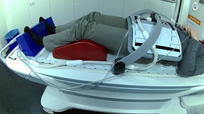
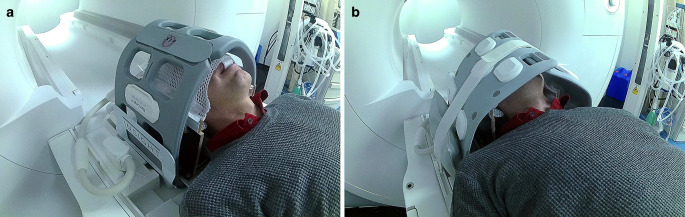
Our novel setup consists of two large 18-channel flexible coils and a specifically designed wooden mask holder mounted on a flat tabletop overlay, which allows patients to be measured in treatment position with mask immobilization. The signal-to-noise ratio (SNR) of this setup was compared to the vendor-provided flexible coil RT setup and the standard setup for diagnostic radiology. The occurrence of motion artifacts was quantified. To develop magnetic resonance imaging (MRI) protocols, we formulated site- and disease-specific clinical objectives.

RESULTS

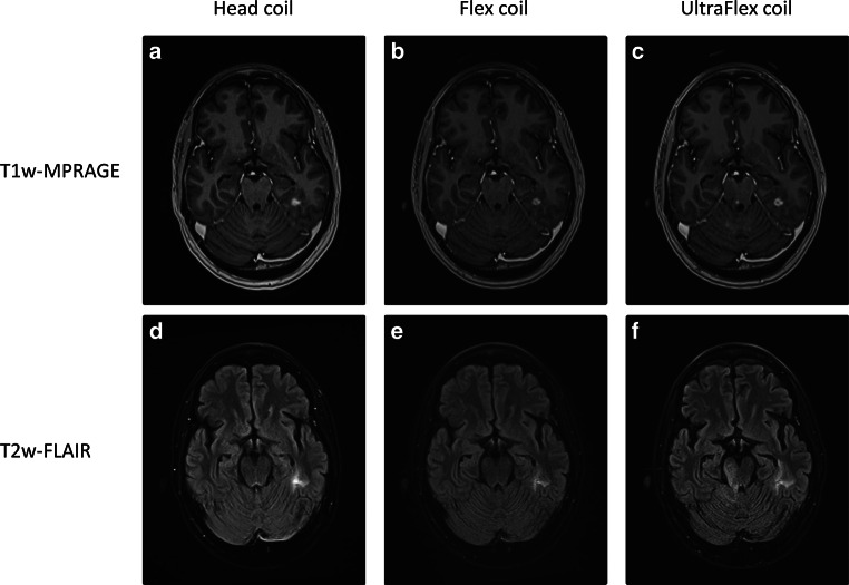
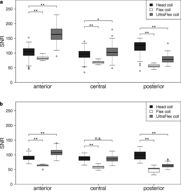
Our novel setup showed mean SNR of 163 ± 28 anteriorly, 104 ± 23 centrally, and 78 ± 14 posteriorly compared to 84 ± 8 and 102 ± 22 anteriorly, 68 ± 6 and 95 ± 20 centrally, and 56 ± 7 and 119 ± 23 posteriorly for the vendor-provided and diagnostic setup, respectively. All differences were significant (p > 0.05). Image quality of our novel setup was judged suitable for contouring by expert-based assessment. Motion artifacts were found in 8/60 patients in the diagnostic setup, whereas none were found for patients in the RT setup. Site-specific core protocols were designed to minimize distortions while optimizing tissue contrast and 3D resolution according to indication-specific objectives.

CONCLUSION

We present a novel setup for high-quality imaging in treatment position that allows use of several immobilization systems enabling MR-only workflows, which could reduce unnecessary dose and registration inaccuracies.

摘要

目的

分享我们在使用新型线圈在治疗位置进行脑成像的基础上,为放射治疗(RT)治疗计划专门安装磁共振(MR)扫描仪的经验,同时介绍为脑和前列腺 RT 治疗计划专门调整的核心序列方案。

材料和方法

我们的新型设备由两个大型 18 通道柔性线圈和一个特定设计的木制面罩固定器组成,安装在一个平板上,使患者在面罩固定下可以在治疗位置进行测量。该设备的信噪比(SNR)与供应商提供的用于 RT 的柔性线圈和用于诊断放射学的标准设备进行了比较。对运动伪影的发生进行了量化。为了开发磁共振成像(MRI)方案,我们制定了基于特定部位和疾病的临床目标。

结果

与供应商提供的和诊断设置相比,我们的新型设备的平均 SNR 分别为 163±28 在前部,104±23 在中央,78±14 在后部,84±8 和 102±22 在前部,68±6 和 95±20 在中央,56±7 和 119±23 在后部。所有差异均有统计学意义(p>0.05)。基于专家评估,新型设备的图像质量被认为适合勾画。在诊断设备中,有 8/60 名患者发现运动伪影,而在 RT 设备中则没有发现运动伪影。根据特定适应症的目标,设计了特定部位的核心方案,以最小化失真,同时优化组织对比度和 3D 分辨率。

结论

我们提出了一种新型的治疗位置高分辨率成像设备,可用于多种固定系统,实现仅使用 MRI 的工作流程,从而减少不必要的剂量和配准不准确。

https://cdn.ncbi.nlm.nih.gov/pmc/blobs/43fe/7892740/04fa6652e997/66_2020_1703_Fig5_HTML.jpg
https://cdn.ncbi.nlm.nih.gov/pmc/blobs/43fe/7892740/c42a0544f0e7/66_2020_1703_Fig1_HTML.jpg
https://cdn.ncbi.nlm.nih.gov/pmc/blobs/43fe/7892740/6f537499c15b/66_2020_1703_Fig2_HTML.jpg
https://cdn.ncbi.nlm.nih.gov/pmc/blobs/43fe/7892740/638c28147aee/66_2020_1703_Fig3_HTML.jpg
https://cdn.ncbi.nlm.nih.gov/pmc/blobs/43fe/7892740/9c681e382aa8/66_2020_1703_Fig4_HTML.jpg
https://cdn.ncbi.nlm.nih.gov/pmc/blobs/43fe/7892740/04fa6652e997/66_2020_1703_Fig5_HTML.jpg

相似文献

1
Implementation of a dedicated 1.5 T MR scanner for radiotherapy treatment planning featuring a novel high-channel coil setup for brain imaging in treatment position.用于放射治疗计划的专用 1.5T MR 扫描仪的实现,具有用于治疗体位下脑成像的新型高通道线圈设置。
Strahlenther Onkol. 2021 Mar;197(3):246-256. doi: 10.1007/s00066-020-01703-y. Epub 2020 Oct 25.
2
Comparison of treatment position with mask immobilization and standard diagnostic setup in intracranial MRI radiotherapy simulation.颅内 MRI 放射治疗模拟中,与面罩固定相比的治疗体位与标准诊断设置的比较。
Strahlenther Onkol. 2021 Jul;197(7):614-621. doi: 10.1007/s00066-021-01776-3. Epub 2021 Apr 21.
3
The influence of patient positioning and immobilization equipment on MR image quality and image registration in radiation therapy.患者体位和固定设备对放射治疗中磁共振图像质量和图像配准的影响。
J Appl Clin Med Phys. 2024 Feb;25(2):e14162. doi: 10.1002/acm2.14162. Epub 2023 Sep 16.
4
Towards integration of PET/MR hybrid imaging into radiation therapy treatment planning.迈向正电子发射断层扫描/磁共振成像(PET/MR)混合成像在放射治疗治疗计划中的整合。
Med Phys. 2014 Jul;41(7):072505. doi: 10.1118/1.4881317.
5
Initial assessment of 3D magnetic resonance fingerprinting (MRF) towards quantitative brain imaging for radiation therapy.3D 磁共振指纹成像(MRF)在放射治疗定量脑成像中的初步评估。
Med Phys. 2020 Mar;47(3):1199-1214. doi: 10.1002/mp.13967. Epub 2019 Dec 30.
6
Comprehensive MRI simulation methodology using a dedicated MRI scanner in radiation oncology for external beam radiation treatment planning.在放射肿瘤学中,使用专用MRI扫描仪进行外照射放疗计划的综合MRI模拟方法。
Med Phys. 2015 Jan;42(1):28-39. doi: 10.1118/1.4896096.
7
Head-and-Neck MRI-only radiotherapy treatment planning: From acquisition in treatment position to pseudo-CT generation.仅使用头部和颈部MRI的放射治疗治疗计划:从在治疗位置进行采集到生成伪CT
Cancer Radiother. 2020 Jul;24(4):288-297. doi: 10.1016/j.canrad.2020.01.008. Epub 2020 Mar 14.
8
Evaluating the image quality of combined positron emission tomography-magnetic resonance images acquired in the pelvic radiotherapy position.评估盆腔放射治疗体位采集的正电子发射断层扫描-磁共振图像的质量。
Phys Med Biol. 2021 Jan 29;66(3):035018. doi: 10.1088/1361-6560/abce1c.
9
Accuracy of MRI-CT registration in brain stereotactic radiotherapy: Impact of MRI acquisition setup and registration method.MRI-CT 配准在脑立体定向放疗中的准确性:MRI 采集设置和配准方法的影响。
Z Med Phys. 2022 Nov;32(4):477-487. doi: 10.1016/j.zemedi.2022.04.004. Epub 2022 May 25.
10
Magnetic resonance imaging for radiotherapy planning of brain cancer patients using immobilization and surface coils.使用固定装置和表面线圈对脑癌患者进行放射治疗计划的磁共振成像。
Phys Med Biol. 2009 Sep 21;54(18):5381-94. doi: 10.1088/0031-9155/54/18/002. Epub 2009 Aug 18.

引用本文的文献

1
MRI distortion correction is associated with improved local control in stereotactic radiotherapy for brain metastases.磁共振成像畸变校正与脑转移瘤立体定向放射治疗中局部控制的改善相关。
Sci Rep. 2025 Mar 17;15(1):9077. doi: 10.1038/s41598-025-93255-3.
2
"sCT-Feasibility" - a feasibility study for deep learning-based MRI-only brain radiotherapy.“sCT-Feasibility”-一项基于深度学习的仅 MRI 脑放疗可行性研究。
Radiat Oncol. 2024 Mar 8;19(1):33. doi: 10.1186/s13014-024-02428-3.
3
Quality requirements for MRI simulation in cranial stereotactic radiotherapy: a guideline from the German Taskforce "Imaging in Stereotactic Radiotherapy".

本文引用的文献

1
Moderately hypofractionated radiotherapy for localized prostate cancer: updated long-term outcome and toxicity analysis.中分割放射治疗局限性前列腺癌:更新的长期结果和毒性分析。
Strahlenther Onkol. 2021 Feb;197(2):124-132. doi: 10.1007/s00066-020-01678-w. Epub 2020 Aug 24.
2
Technological quality requirements for stereotactic radiotherapy : Expert review group consensus from the DGMP Working Group for Physics and Technology in Stereotactic Radiotherapy.立体定向放射治疗的技术质量要求:来自立体定向放射治疗物理与技术工作组的 DGMP 专家组共识。
Strahlenther Onkol. 2020 May;196(5):421-443. doi: 10.1007/s00066-020-01583-2. Epub 2020 Mar 24.
3
头部立体定向放射治疗中 MRI 模拟的质量要求:德国“立体定向放射治疗中的影像学”工作组指南。
Strahlenther Onkol. 2024 Jan;200(1):1-18. doi: 10.1007/s00066-023-02183-6. Epub 2024 Jan 2.
4
The influence of patient positioning and immobilization equipment on MR image quality and image registration in radiation therapy.患者体位和固定设备对放射治疗中磁共振图像质量和图像配准的影响。
J Appl Clin Med Phys. 2024 Feb;25(2):e14162. doi: 10.1002/acm2.14162. Epub 2023 Sep 16.
5
Impact of magnetic resonance imaging-related geometric distortion of dose distribution in fractionated stereotactic radiotherapy in patients with brain metastases.磁共振成像相关的分次立体定向放疗中脑转移瘤剂量分布的几何变形的影响。
Strahlenther Onkol. 2024 Jan;200(1):39-48. doi: 10.1007/s00066-023-02120-7. Epub 2023 Aug 17.
6
Synthetic CTs for MRI-only brain RT treatment: integration of immobilization systems.用于仅 MRI 脑 RT 治疗的合成 CT:固定系统的整合。
Strahlenther Onkol. 2023 Aug;199(8):739-748. doi: 10.1007/s00066-023-02090-w. Epub 2023 Jun 7.
7
Quality assurance and temporal stability of a 1.5 T MRI scanner for MR-guided Photon and Particle Therapy.用于磁共振引导光子和粒子治疗的1.5T磁共振成像扫描仪的质量保证和时间稳定性
Z Med Phys. 2025 May;35(2):204-217. doi: 10.1016/j.zemedi.2023.04.004. Epub 2023 May 5.
8
Usability of magnetic resonance images acquired at a novel low-field 0.55 T scanner for brain radiotherapy treatment planning.新型0.55T低场强扫描仪获取的磁共振图像在脑放射治疗计划中的可用性。
Phys Imaging Radiat Oncol. 2023 Jan 13;25:100412. doi: 10.1016/j.phro.2023.100412. eCollection 2023 Jan.
9
Prospective Evaluation of CD45RA+/CCR7- Effector Memory T (T) Cell Subsets in Patients with Primary and Secondary Brain Tumors during Radiotherapy of the Brain within the Scope of the Prospective Glio-CMV-01 Clinical Trial.原发性和继发性脑肿瘤患者在脑放射治疗中前瞻性 Glio-CMV-01 临床试验范围内的 CD45RA+/CCR7-效应记忆 T(T)细胞亚群的前瞻性评估。
Cells. 2023 Feb 4;12(4):516. doi: 10.3390/cells12040516.
10
Clinical implementation of magnetic resonance imaging simulation for radiation oncology planning: 5 year experience.磁共振成像模拟在放射肿瘤计划中的临床应用:5 年经验。
Radiat Oncol. 2023 Feb 7;18(1):27. doi: 10.1186/s13014-023-02209-4.
Magnetic resonance imaging for brain stereotactic radiotherapy : A review of requirements and pitfalls.
磁共振成像在脑立体定向放疗中的应用:需求与误区综述。
Strahlenther Onkol. 2020 May;196(5):444-456. doi: 10.1007/s00066-020-01604-0. Epub 2020 Mar 23.
4
Brain and Head-and-Neck MRI in Immobilization Mask: A Practical Solution for MR-Only Radiotherapy.使用固定面罩进行脑部和头颈部MRI:仅使用MRI进行放射治疗的实用解决方案。
Front Oncol. 2019 Jul 17;9:647. doi: 10.3389/fonc.2019.00647. eCollection 2019.
5
Brain Tumor-Enhancement Visualization and Morphometric Assessment: A Comparison of MPRAGE, SPACE, and VIBE MRI Techniques.脑肿瘤增强可视化与形态计量评估:MPRAGE、SPACE 和 VIBE MRI 技术的比较。
AJNR Am J Neuroradiol. 2019 Jul;40(7):1140-1148. doi: 10.3174/ajnr.A6096. Epub 2019 Jun 20.
6
MRI-guided localization of the dominant intraprostatic lesion and dose analysis of volumetric modulated arc therapy planning for prostate cancer.MRI 引导下前列腺内优势病灶的定位和前列腺癌容积调强弧形治疗计划的剂量分析。
Strahlenther Onkol. 2019 Feb;195(2):145-152. doi: 10.1007/s00066-018-1364-5. Epub 2018 Sep 12.
7
Dose evaluation of fast synthetic-CT generation using a generative adversarial network for general pelvis MR-only radiotherapy.使用生成对抗网络进行快速合成 CT 生成的剂量评估,用于普通骨盆仅磁共振放疗。
Phys Med Biol. 2018 Sep 10;63(18):185001. doi: 10.1088/1361-6560/aada6d.
8
MR-Only Brain Radiation Therapy: Dosimetric Evaluation of Synthetic CTs Generated by a Dilated Convolutional Neural Network.仅行磁共振脑放疗:利用扩张卷积神经网络生成的合成 CT 的剂量学评估。
Int J Radiat Oncol Biol Phys. 2018 Nov 15;102(4):801-812. doi: 10.1016/j.ijrobp.2018.05.058. Epub 2018 Jun 4.
9
Magnetic Resonance Imaging for Target Delineation and Daily Treatment Modification.磁共振成像在靶区勾画和日常治疗修正中的应用。
Semin Radiat Oncol. 2018 Jun;28(3):178-184. doi: 10.1016/j.semradonc.2018.02.002.
10
Improvements in High Resolution Laryngeal Magnetic Resonance Imaging for Preoperative Transoral Laser Microsurgery and Radiotherapy Considerations in Early Lesions.早期病变术前经口激光显微手术及放射治疗相关的高分辨率喉部磁共振成像的改进
Front Oncol. 2018 Jun 6;8:216. doi: 10.3389/fonc.2018.00216. eCollection 2018.