University of Houston, College of Optometry, Houston, Texas, United States.
School of Optometry and Vision Science, University of New South Wales, Sydney, Australia.
Invest Ophthalmol Vis Sci. 2020 Oct 1;61(12):26. doi: 10.1167/iovs.61.12.26.
Decreased corneal nerve fiber density and higher corneal epithelial dendritic cells have been reported in established patients with type 2 diabetes; however, alterations in the subbasal nerve plexus in prediabetes with healthy subjects or subjects with diabetes is limited. The study aimed to determine corneal nerve fiber density and morphology and dendritic cell density between healthy subjects and those with prediabetes or type 2 diabetes.
Fifty-two subjects (aged 30-70 years) were recruited. Blood samples and body metrics were taken. Subjects were grouped as: healthy controls (hemoglobin A1c [HbA1c] < 5.7%), prediabetes (5.7-6.4%), and type 2 diabetes (> 6.4% or physician diagnosis). Central corneal subbasal nerve plexus was imaged using in vivo confocal microscopy. Corneal nerve fiber density and morphology, including interconnections and tortuosity, and dendritic cell density were assessed. Kruskal-Wallis tests were carried out to compare differences in the examined variables between groups. Spearman correlations were carried out to examine the associations between body metrics with HbA1c and corneal findings.
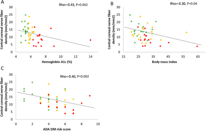
Seventeen healthy controls, 20 subjects with prediabetes, and 15 subjects with type 2 diabetes completed this study. Central corneal nerve fiber density was significantly lower in type 2 diabetes compared to prediabetes (P = 0.045) and healthy controls (P = 0.001). No differences were found in central corneal nerve fiber interconnections, tortuosity, or dendritic cell density between groups. There was a significant association between HbA1c and corneal nerve fiber density (rho = -0.45, P = 0.001) and body mass index (BMI; rho = -0.30, P = 0.04).
Increased HbA1c values are associated with decreased corneal nerve fiber density across the spectrum of type 2 diabetes.
有报道称,2 型糖尿病患者的角膜神经纤维密度降低,角膜上皮树突状细胞增多;然而,糖尿病前期患者与健康受试者或糖尿病患者的基底神经丛改变有限。本研究旨在确定健康受试者与糖尿病前期或 2 型糖尿病患者之间的角膜神经纤维密度和形态以及树突状细胞密度。
招募了 52 名(年龄 30-70 岁)受试者。采集血样和身体指标。受试者分为三组:健康对照组(糖化血红蛋白(HbA1c)<5.7%)、糖尿病前期组(5.7-6.4%)和 2 型糖尿病组(>6.4%或医生诊断)。使用活体共聚焦显微镜对中央角膜基底神经丛进行成像。评估角膜神经纤维密度和形态,包括神经纤维的相互连接和弯曲程度,以及树突状细胞密度。采用 Kruskal-Wallis 检验比较组间各变量的差异。采用 Spearman 相关分析评估身体指标与 HbA1c 及角膜检查结果之间的相关性。
本研究共纳入 17 名健康对照组、20 名糖尿病前期组和 15 名 2 型糖尿病组受试者。与糖尿病前期组和健康对照组相比,2 型糖尿病组的中央角膜神经纤维密度明显降低(P=0.045 和 P=0.001)。各组间中央角膜神经纤维的相互连接、弯曲程度或树突状细胞密度无差异。HbA1c 与角膜神经纤维密度呈显著负相关(rho=-0.45,P=0.001),与体重指数(BMI)呈显著负相关(rho=-0.30,P=0.04)。
HbA1c 值升高与 2 型糖尿病患者角膜神经纤维密度降低有关。