Yuan Yusong, Li Dongdong, Yu Fei, Kang Xuejing, Xu Hailin, Zhang Peixun
Department of Trauma and Orthopedics, Peking University People's Hospital, Peking University, Beijing, China.
Key Laboratory of Trauma and Neural Regeneration, Ministry of Education, Peking University, Beijing, China.
Front Neurosci. 2020 Sep 29;14:565870. doi: 10.3389/fnins.2020.565870. eCollection 2020.
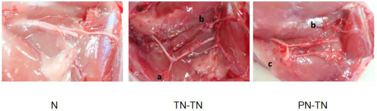
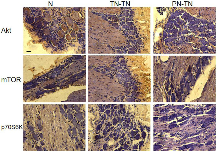
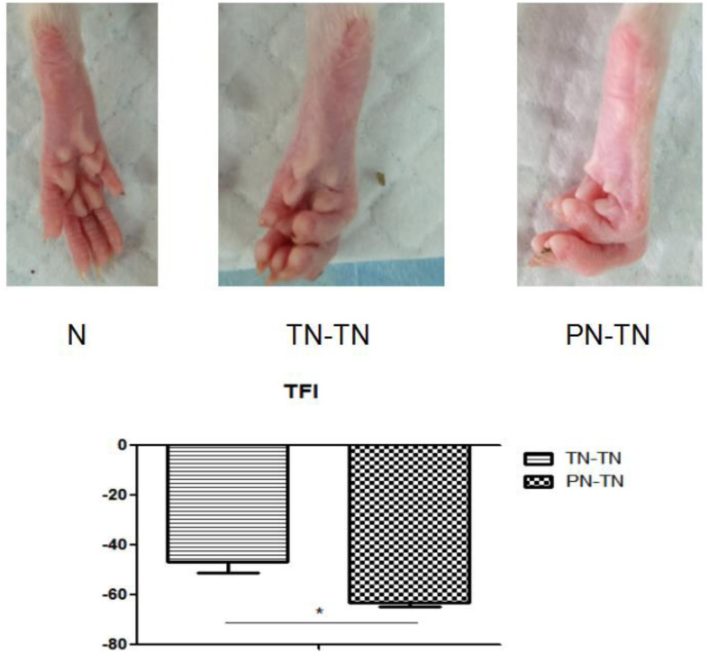
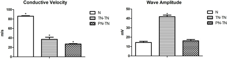
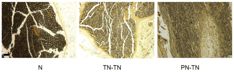
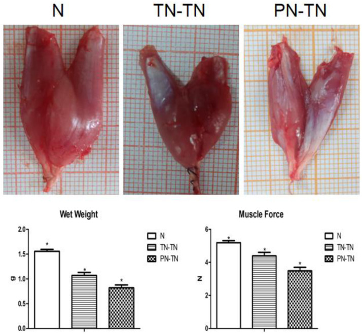
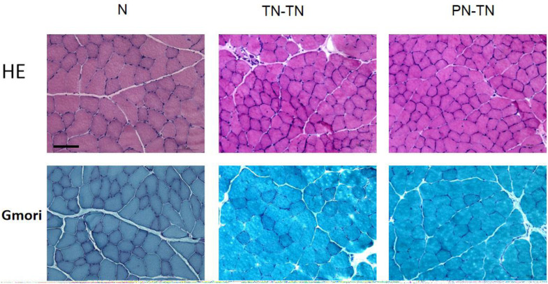
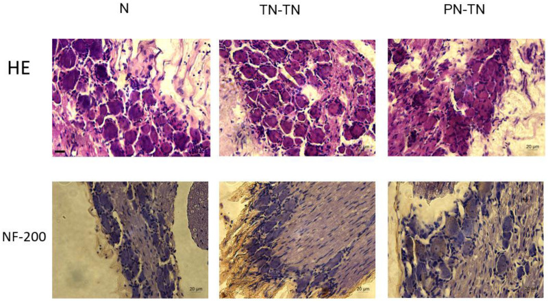
Peripheral nerve injury repair has been considered a difficult problem in the field of trauma for a long time. Conventional surgical methods are not applicable in some special types of nerve injury, prompting scholars to seek to develop more effective nerve translocation repair technologies. The purpose of this study was to explore the functional state of neurons in injured lower limbs after translocation repair, with a view to preliminarily clarify the molecular mechanisms underlying this process. Eighteen Sprague-Dawley rats were divided into the normal, tibial nerve repair, and common peroneal nerve transposition repair tibial nerve groups. Nerve function assessment and immunohistochemical staining of neurofilament 200 (NF-200), protein kinase B (Akt), mammalian target of rapamycin (mTOR), and ribosomal protein S6 kinase (p70S6K) in the dorsal root ganglia were performed at 12 weeks after surgery. Tibial nerve function and neuroelectrophysiological analysis, osmic acid staining, muscle strength testing, and muscle fiber staining showed that the nerve translocation repair could restore the function of the recipient nerve to a certain extent; however, the repair was not as efficient as the repair. Immunohistochemical staining showed that the translocation repair resulted in changes in the microstructure of neuronal cell bodies, and the expressions of Akt, mTOR, and p70S6K in the three dorsal root ganglia groups were significantly different ( < 0.05). This study demonstrates that the nerve translocation repair technology sets up a new reflex loop, with the corresponding neuroskeletal adjustments, in which, donor neurons dominate the recipient nerves. This indicates that nerve translocation repair technology can lead to neuronal remodeling and is important as a supplementary treatment for a peripheral nerve injury. Furthermore, the Akt/mTOR/p70S6K signaling pathway may be involved in the formation of the new neural reflex loop created as a result of the translocation repair.
长期以来,周围神经损伤修复一直被认为是创伤领域的一个难题。传统手术方法不适用于某些特殊类型的神经损伤,这促使学者们寻求开发更有效的神经移位修复技术。本研究的目的是探讨移位修复后损伤下肢神经元的功能状态,以期初步阐明这一过程的分子机制。将18只Sprague-Dawley大鼠分为正常组、胫神经修复组和腓总神经移位修复胫神经组。术后12周进行神经功能评估以及背根神经节中神经丝200(NF-200)、蛋白激酶B(Akt)、雷帕霉素哺乳动物靶点(mTOR)和核糖体蛋白S6激酶(p70S6K)的免疫组织化学染色。胫神经功能及神经电生理分析、锇酸染色、肌力测试和肌纤维染色结果显示,神经移位修复可在一定程度上恢复受区神经功能;然而,修复效果不如直接修复。免疫组织化学染色显示,移位修复导致神经元细胞体微观结构发生变化,三个背根神经节组中Akt、mTOR和p70S6K的表达存在显著差异(P<0.05)。本研究表明,神经移位修复技术建立了一个新的反射环路,并伴有相应的神经骨骼调整,其中供体神经元支配受区神经。这表明神经移位修复技术可导致神经元重塑,作为周围神经损伤的一种辅助治疗具有重要意义。此外,Akt/mTOR/p70S6K信号通路可能参与了移位修复所形成的新神经反射环路的形成。