• 文献检索
  • 文档翻译
  • 深度研究
  • 学术资讯
  • Suppr Zotero 插件Zotero 插件
  • 邀请有礼
  • 套餐&价格
  • 历史记录
应用&插件
Suppr Zotero 插件Zotero 插件浏览器插件Mac 客户端Windows 客户端微信小程序
定价
高级版会员购买积分包购买API积分包
服务
文献检索文档翻译深度研究API 文档MCP 服务
关于我们
关于 Suppr公司介绍联系我们用户协议隐私条款
关注我们

Suppr 超能文献

核心技术专利:CN118964589B侵权必究
粤ICP备2023148730 号-1Suppr @ 2026

文献检索

告别复杂PubMed语法,用中文像聊天一样搜索,搜遍4000万医学文献。AI智能推荐,让科研检索更轻松。

立即免费搜索

文件翻译

保留排版,准确专业,支持PDF/Word/PPT等文件格式,支持 12+语言互译。

免费翻译文档

深度研究

AI帮你快速写综述,25分钟生成高质量综述,智能提取关键信息,辅助科研写作。

立即免费体验

WNT11 条件培养基通过非经典 WNT-PKC-JNK 信号通路激活促进血管生成。

WNT11-Conditioned Medium Promotes Angiogenesis through the Activation of Non-Canonical WNT-PKC-JNK Signaling Pathway.

机构信息

Department of Pathology and Laboratory Medicine, University of Cincinnati Medical Center, Cincinnati, OH 45267, USA.

出版信息

Genes (Basel). 2020 Oct 29;11(11):1277. doi: 10.3390/genes11111277.

DOI:10.3390/genes11111277
PMID:33137935
原文链接:https://pmc.ncbi.nlm.nih.gov/articles/PMC7694138/
Abstract

BACKGROUND

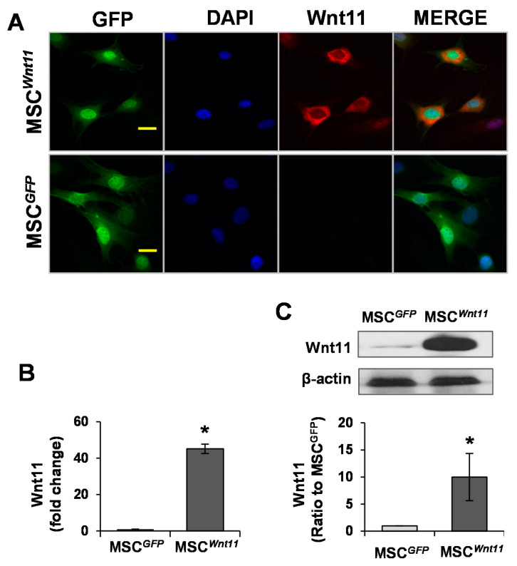
We demonstrated that the transduction of Wnt11 into mesenchymal stem cells (MSCs) (MSC) promotes these cells differentiation into cardiac phenotypes. In the present study, we investigated the paracrine effects of MSC on cardiac function and angiogenesis.

METHODS AND RESULTS

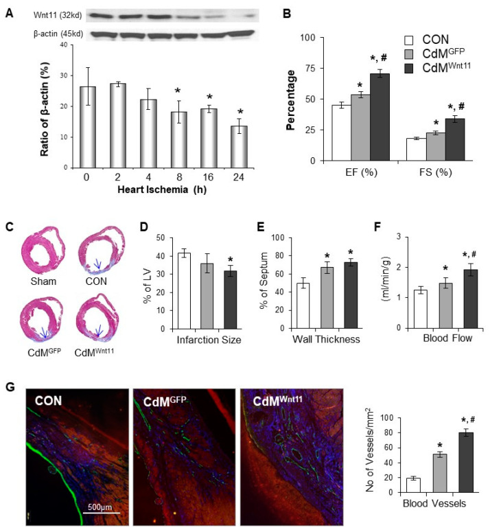
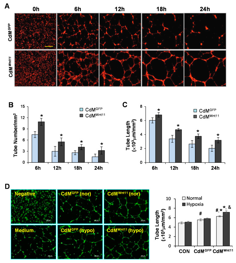
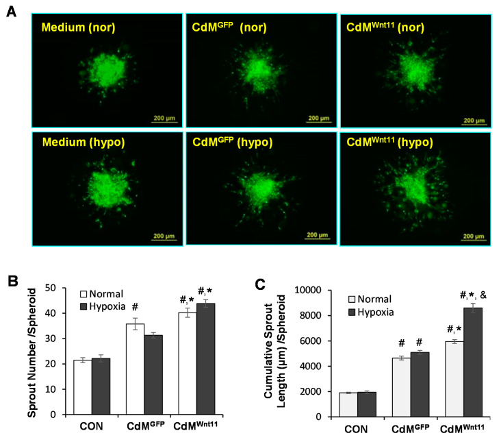
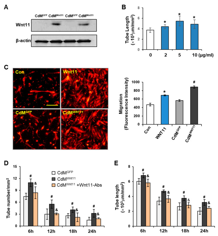
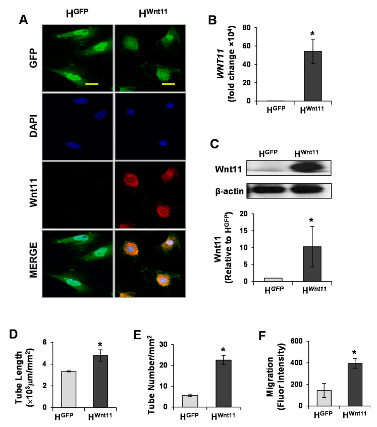
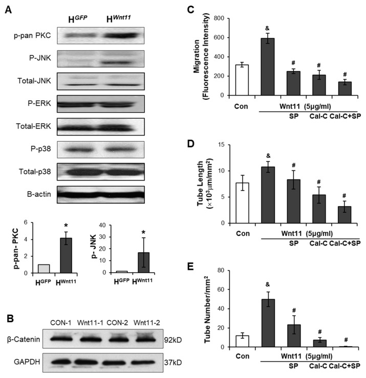
Conditioned medium was collected from MSC (CdM) and their control cells (CdM). CdM, especially obtained from MSC exposed to hypoxia, significantly promoted human umbilical vein endothelial cells (HUVECs) migration and increased capillary-like tube (CLT) formation, which was blocked by Wnt11 neutralizing antibody. Wnt11 protein was significantly higher in CdM compared to that in CdM. Directly treating HUVECs with recombinant Wnt11 protein significantly increased CLT formation, which was abrogated by treating cells with the JNK inhibitor SP600125, as well as the PKC inhibitor Calphostin-C. Moreover, the transfection of Wnt11 to HUVECs (H) significantly increased CLT formation and HUVEC migration, as well as upregulated p-pan-PKC and p-JNK expression. Injection of CdM into the peri-infarct region in a rat acute myocardial infarction (AMI) model significantly improved cardiac function, reduced infarct size, and increased myocardial blood flow and blood vessel density in the ischemic area.

CONCLUSION

Wnt11 released from MSC increased angiogenesis and improved cardiac function via non-canonical Wnt-PKC-JNK dependent pathways.

摘要

背景

我们证明了将 Wnt11 转导到间充质干细胞(MSC)中可促进这些细胞向心脏表型分化。在本研究中,我们研究了 MSC 对心脏功能和血管生成的旁分泌作用。

方法和结果

收集 MSC(CdM)及其对照细胞(CdM)的条件培养基。CdM,特别是来自暴露于缺氧条件下的 MSC 的 CdM,可显著促进人脐静脉内皮细胞(HUVEC)的迁移,并增加毛细血管样管(CLT)的形成,而 Wnt11 中和抗体可阻断该作用。与 CdM 相比,CdM 中的 Wnt11 蛋白明显更高。直接用重组 Wnt11 蛋白处理 HUVECs 可显著增加 CLT 的形成,而用 JNK 抑制剂 SP600125 和 PKC 抑制剂 Calphostin-C 处理细胞可消除这种作用。此外,Wnt11 转染到 HUVECs(H)中可显著增加 CLT 的形成和 HUVEC 的迁移,并上调 p-pan-PKC 和 p-JNK 的表达。将 CdM 注射到大鼠急性心肌梗死(AMI)模型的梗死周边区可显著改善心脏功能,减少梗死面积,并增加缺血区的心肌血流和血管密度。

结论

MSC 释放的 Wnt11 通过非经典 Wnt-PKC-JNK 依赖途径增加血管生成并改善心脏功能。

https://cdn.ncbi.nlm.nih.gov/pmc/blobs/368f/7694138/d315f99c8e75/genes-11-01277-g007.jpg
https://cdn.ncbi.nlm.nih.gov/pmc/blobs/368f/7694138/19ae50361e78/genes-11-01277-g001.jpg
https://cdn.ncbi.nlm.nih.gov/pmc/blobs/368f/7694138/9bd4c30664c8/genes-11-01277-g002.jpg
https://cdn.ncbi.nlm.nih.gov/pmc/blobs/368f/7694138/adc8aaf95082/genes-11-01277-g003.jpg
https://cdn.ncbi.nlm.nih.gov/pmc/blobs/368f/7694138/0f9be7eb8005/genes-11-01277-g004.jpg
https://cdn.ncbi.nlm.nih.gov/pmc/blobs/368f/7694138/712373353946/genes-11-01277-g005.jpg
https://cdn.ncbi.nlm.nih.gov/pmc/blobs/368f/7694138/59315f7831a5/genes-11-01277-g006.jpg
https://cdn.ncbi.nlm.nih.gov/pmc/blobs/368f/7694138/d315f99c8e75/genes-11-01277-g007.jpg

相似文献

1
WNT11-Conditioned Medium Promotes Angiogenesis through the Activation of Non-Canonical WNT-PKC-JNK Signaling Pathway.WNT11 条件培养基通过非经典 WNT-PKC-JNK 信号通路激活促进血管生成。
Genes (Basel). 2020 Oct 29;11(11):1277. doi: 10.3390/genes11111277.
2
Paracrine effect of Wnt11-overexpressing mesenchymal stem cells on ischemic injury.Wnt11 过表达间充质干细胞的旁分泌作用对缺血性损伤的影响。
Stem Cells Dev. 2012 Mar 1;21(4):598-608. doi: 10.1089/scd.2011.0071. Epub 2011 Jun 1.
3
Paracrine factors released by GATA-4 overexpressed mesenchymal stem cells increase angiogenesis and cell survival.过表达 GATA-4 的间充质干细胞释放的旁分泌因子可增加血管生成和细胞存活。
Am J Physiol Heart Circ Physiol. 2010 Dec;299(6):H1772-81. doi: 10.1152/ajpheart.00557.2010. Epub 2010 Sep 24.
4
Wnt11 preserves mitochondrial membrane potential and protects cardiomyocytes against hypoxia through paracrine signaling.Wnt11通过旁分泌信号传导维持线粒体膜电位并保护心肌细胞免受缺氧损伤。
J Cell Biochem. 2020 Feb;121(2):1144-1155. doi: 10.1002/jcb.29349. Epub 2019 Aug 28.
5
Transduction of Wnt11 promotes mesenchymal stem cell transdifferentiation into cardiac phenotypes.Wnt11 的转导促进间充质干细胞向心脏表型的转分化。
Stem Cells Dev. 2011 Oct;20(10):1771-8. doi: 10.1089/scd.2010.0380. Epub 2011 Feb 26.
6
Co-expression of Akt1 and Wnt11 promotes the proliferation and cardiac differentiation of mesenchymal stem cells and attenuates hypoxia/reoxygenation-induced cardiomyocyte apoptosis.Akt1 和 Wnt11 的共表达促进间充质干细胞的增殖和心脏分化,并减轻低氧/复氧诱导的心肌细胞凋亡。
Biomed Pharmacother. 2018 Dec;108:508-514. doi: 10.1016/j.biopha.2018.09.047. Epub 2018 Sep 19.
7
Heparanase released from mesenchymal stem cells activates integrin beta1/HIF-2alpha/Flk-1 signaling and promotes endothelial cell migration and angiogenesis.间充质干细胞释放的乙酰肝素酶激活整合素β1/HIF-2α/Flk-1信号通路并促进内皮细胞迁移和血管生成。
Stem Cells. 2015 Jun;33(6):1850-1862. doi: 10.1002/stem.1995.
8
Non-canonical Wnt signaling enhances differentiation of human circulating progenitor cells to cardiomyogenic cells.非经典Wnt信号通路增强人循环祖细胞向心肌生成细胞的分化。
J Biol Chem. 2005 Apr 29;280(17):16838-42. doi: 10.1074/jbc.M500323200. Epub 2005 Feb 8.
9
Co-culture of mesenchymal stem cells with umbilical vein endothelial cells under hypoxic condition.间充质干细胞与脐静脉内皮细胞在缺氧条件下的共培养。
J Huazhong Univ Sci Technolog Med Sci. 2012 Apr;32(2):173-180. doi: 10.1007/s11596-012-0031-9. Epub 2012 Apr 20.
10
MicroRNA-377 regulates mesenchymal stem cell-induced angiogenesis in ischemic hearts by targeting VEGF.微小RNA-377通过靶向血管内皮生长因子调控缺血心脏中间充质干细胞诱导的血管生成。
PLoS One. 2014 Sep 24;9(9):e104666. doi: 10.1371/journal.pone.0104666. eCollection 2014.

引用本文的文献

1
Wnt Signaling Inhibitors as Therapeutic Approach in Ischemic Heart Disease.Wnt信号通路抑制剂作为缺血性心脏病的治疗方法
Molecules. 2024 Dec 17;29(24):5958. doi: 10.3390/molecules29245958.
2
Structural and Functional Insights into Dishevelled-Mediated Wnt Signaling.深入了解蓬乱蛋白介导的 Wnt 信号传导的结构与功能。
Cells. 2024 Nov 11;13(22):1870. doi: 10.3390/cells13221870.
3
Unveiling the Potential of Migrasomes: A Machine-Learning-Driven Signature for Diagnosing Acute Myocardial Infarction.揭示迁移小体的潜力:一种机器学习驱动的急性心肌梗死诊断标志物

本文引用的文献

1
Bone marrow-derived mesenchymal stromal cell treatment in patients with ischaemic heart failure: final 4-year follow-up of the MSC-HF trial.缺血性心力衰竭患者的骨髓间充质基质细胞治疗:MSC-HF试验的4年最终随访
Eur J Heart Fail. 2020 May;22(5):884-892. doi: 10.1002/ejhf.1700. Epub 2019 Dec 21.
2
Intramyocardial Bone Marrow Stem Cells in Patients Undergoing Cardiac Surgical Revascularization.心肌内骨髓干细胞在接受心脏外科血运重建术的患者中的应用。
Ann Thorac Surg. 2020 Apr;109(4):1142-1149. doi: 10.1016/j.athoracsur.2019.07.093. Epub 2019 Sep 14.
3
Ascl1-induced Wnt11 regulates neuroendocrine differentiation, cell proliferation, and E-cadherin expression in small-cell lung cancer and Wnt11 regulates small-cell lung cancer biology.
Biomedicines. 2024 Jul 22;12(7):1626. doi: 10.3390/biomedicines12071626.
4
Therapeutic potential of mesenchymal stem cells for cerebral small vessel disease.间充质干细胞对脑小血管病的治疗潜力
Regen Ther. 2024 Feb 22;25:377-386. doi: 10.1016/j.reth.2023.11.002. eCollection 2024 Mar.
5
Ischemia-reperfusion injury: molecular mechanisms and therapeutic targets.缺血再灌注损伤:分子机制与治疗靶点。
Signal Transduct Target Ther. 2024 Jan 8;9(1):12. doi: 10.1038/s41392-023-01688-x.
6
Wilforine inhibits rheumatoid arthritis pathology through the Wnt11/β-catenin signaling pathway axis.狼毒素通过 Wnt11/β-catenin 信号通路轴抑制类风湿关节炎病理。
Arthritis Res Ther. 2023 Dec 14;25(1):243. doi: 10.1186/s13075-023-03224-2.
7
Cardiac stem cells: Current knowledge and future prospects.心脏干细胞:当前认知与未来展望。
World J Stem Cells. 2022 Jan 26;14(1):1-40. doi: 10.4252/wjsc.v14.i1.1.
8
Cardiac Wnt5a and Wnt11 promote fibrosis by the crosstalk of FZD5 and EGFR signaling under pressure overload.心脏 Wnt5a 和 Wnt11 在压力超负荷下通过 FZD5 和 EGFR 信号的串扰促进纤维化。
Cell Death Dis. 2021 Sep 25;12(10):877. doi: 10.1038/s41419-021-04152-2.
9
Emerging Trends in Deciphering the Pathogenesis of Human Diseases through Genetic Analysis.通过基因分析揭示人类疾病发病机制的新兴趋势。
Genes (Basel). 2021 Jan 13;12(1):96. doi: 10.3390/genes12010096.
Ascl1 诱导的 Wnt11 调节小细胞肺癌的神经内分泌分化、细胞增殖和 E-钙黏蛋白表达,Wnt11 调节小细胞肺癌生物学。
Lab Invest. 2019 Nov;99(11):1622-1635. doi: 10.1038/s41374-019-0277-y. Epub 2019 Jun 23.
4
Reactivating endogenous mechanisms of cardiac regeneration via paracrine boosting using the human amniotic fluid stem cell secretome.通过使用人羊膜间充质干细胞 secretome 的旁分泌增强作用来激活心脏再生的内源性机制。
Int J Cardiol. 2019 Jul 15;287:87-95. doi: 10.1016/j.ijcard.2019.04.011. Epub 2019 Apr 4.
5
Induced pluripotent stem cell-conditional medium inhibits H9C2 cardiomyocytes apoptosis via autophagy flux and Wnt/β-catenin pathway.诱导多能干细胞条件培养基通过自噬流和 Wnt/β-连环蛋白通路抑制 H9C2 心肌细胞凋亡。
J Cell Mol Med. 2019 Jun;23(6):4358-4374. doi: 10.1111/jcmm.14327. Epub 2019 Apr 7.
6
Wnt/β-catenin-mediated signaling re-activates proliferation of matured cardiomyocytes.Wnt/β-catenin 信号通路介导成熟心肌细胞的增殖。
Stem Cell Res Ther. 2018 Dec 7;9(1):338. doi: 10.1186/s13287-018-1086-8.
7
Co-expression of Akt1 and Wnt11 promotes the proliferation and cardiac differentiation of mesenchymal stem cells and attenuates hypoxia/reoxygenation-induced cardiomyocyte apoptosis.Akt1 和 Wnt11 的共表达促进间充质干细胞的增殖和心脏分化,并减轻低氧/复氧诱导的心肌细胞凋亡。
Biomed Pharmacother. 2018 Dec;108:508-514. doi: 10.1016/j.biopha.2018.09.047. Epub 2018 Sep 19.
8
Comparative study of regenerative effects of mesenchymal stem cells derived from placental amnion, chorion and umbilical cord on dermal wounds.胎盘羊膜、绒毛膜和脐带间充质干细胞对皮肤创面再生作用的比较研究。
Placenta. 2018 May;65:37-46. doi: 10.1016/j.placenta.2018.04.004. Epub 2018 Apr 10.
9
Effects of Endothelial and Mesenchymal Stem Cells on Improving Myocardial Function in a Sheep Animal Model.内皮细胞和间充质干细胞对绵羊动物模型心肌功能改善的作用
J Tehran Heart Cent. 2017 Apr;12(2):65-71.
10
Wnt ligand presentation and reception: from the stem cell niche to tissue engineering.Wnt 配体的呈递和接受:从干细胞龛到组织工程。
Open Biol. 2017 Aug;7(8). doi: 10.1098/rsob.170140.