Thayse Kathleen, Kindt Nadège, Laurent Sophie, Carlier Stéphane
Laboratory of Cardiology, Faculty of Medicine and Pharmacy, Université de Mons, 7000 Mons, Belgium.
Laboratory of Oncology and Experimental Surgery, Institut Jules Bordet, Université Libre de Bruxelles, 1000 Brussels, Belgium.
Biology (Basel). 2020 Oct 29;9(11):368. doi: 10.3390/biology9110368.
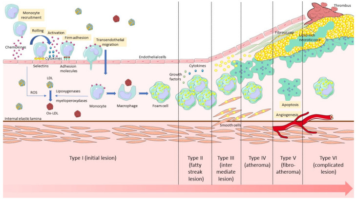
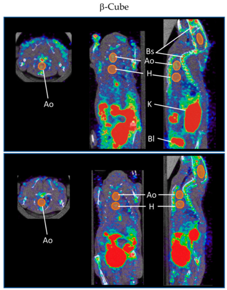
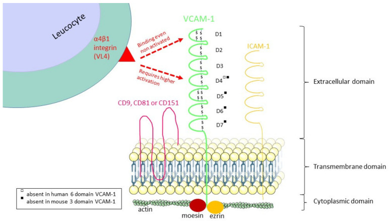
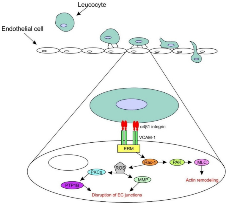
Atherosclerosis is a progressive chronic arterial disease characterised by atheromatous plaque formation in the intima of the arterial wall. Several invasive and non-invasive imaging techniques have been developed to detect and characterise atherosclerosis in the vessel wall: anatomic/structural imaging, functional imaging and molecular imaging. In molecular imaging, vascular cell adhesion molecule-1 (VCAM-1) is a promising target for the non-invasive detection of atherosclerosis and for the assessment of novel antiatherogenic treatments. VCAM-1 is an adhesion molecule expressed on the activated endothelial surface that binds leucocyte ligands and therefore promotes leucocyte adhesion and transendothelial migration. Hence, for several years, there has been an increase in molecular imaging methods for detecting VCAM-1 in MRI, PET, SPECT, optical imaging and ultrasound. The use of microparticles of iron oxide (MPIO), ultrasmall superparamagnetic iron oxide (USPIO), microbubbles, echogenic immunoliposomes, peptides, nanobodies and other nanoparticles has been described. However, these approaches have been tested in animal models, and the remaining challenge is bench-to-bedside development and clinical applicability.
动脉粥样硬化是一种进行性慢性动脉疾病,其特征是动脉壁内膜形成动脉粥样斑块。已经开发了几种有创和无创成像技术来检测和表征血管壁中的动脉粥样硬化:解剖/结构成像、功能成像和分子成像。在分子成像中,血管细胞粘附分子-1(VCAM-1)是动脉粥样硬化无创检测和新型抗动脉粥样硬化治疗评估的一个有前景的靶点。VCAM-1是一种在活化内皮表面表达的粘附分子,它结合白细胞配体,从而促进白细胞粘附和跨内皮迁移。因此,多年来,用于在MRI、PET、SPECT、光学成像和超声中检测VCAM-1的分子成像方法不断增加。已经描述了使用氧化铁微粒(MPIO)、超小超顺磁性氧化铁(USPIO)、微泡、回声免疫脂质体、肽、纳米抗体和其他纳米颗粒。然而,这些方法已在动物模型中进行了测试,但剩下的挑战是从实验室到临床的开发和临床适用性。