Division of Cardiology Mount Sinai Hospital, Sinai Health Toronto Ontario Canada.
Faculty of Kinesiology and Physical Education University of Toronto Ontario Canada.
J Am Heart Assoc. 2020 Nov 17;9(22):e016339. doi: 10.1161/JAHA.120.016339. Epub 2020 Nov 6.
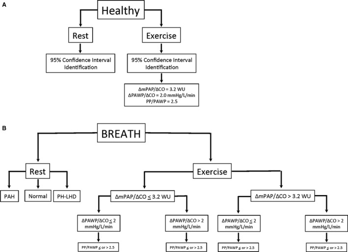
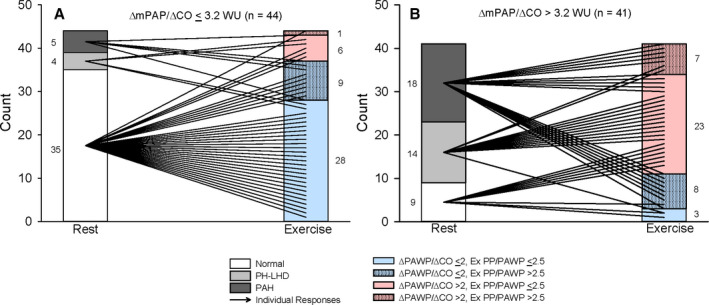
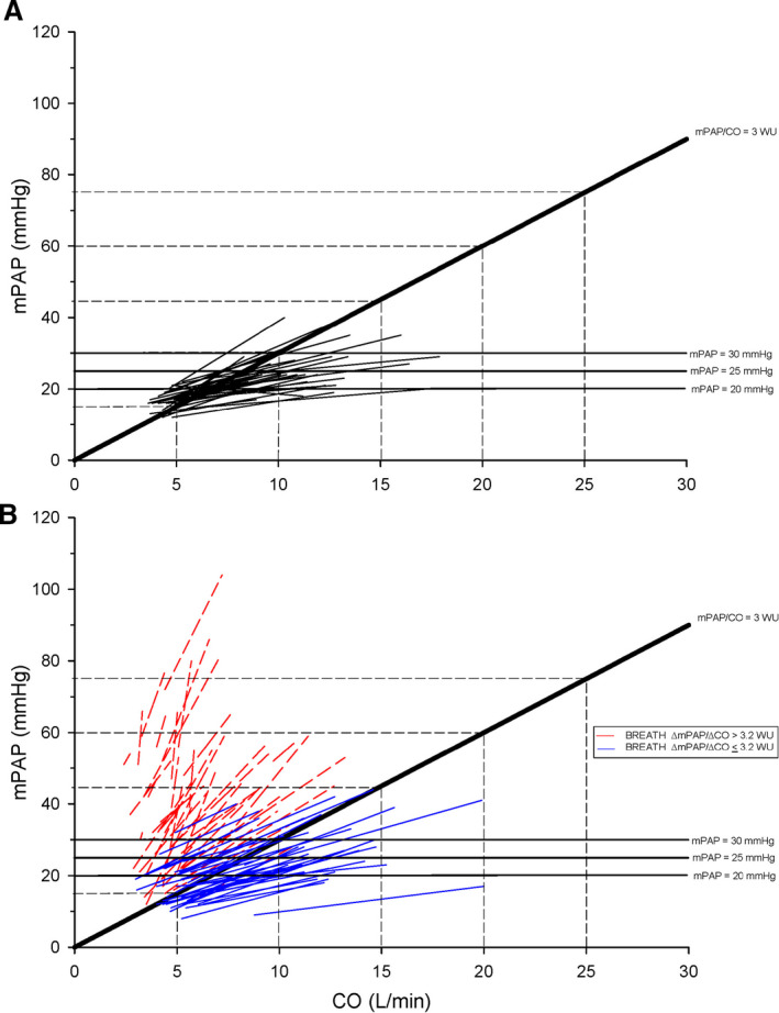
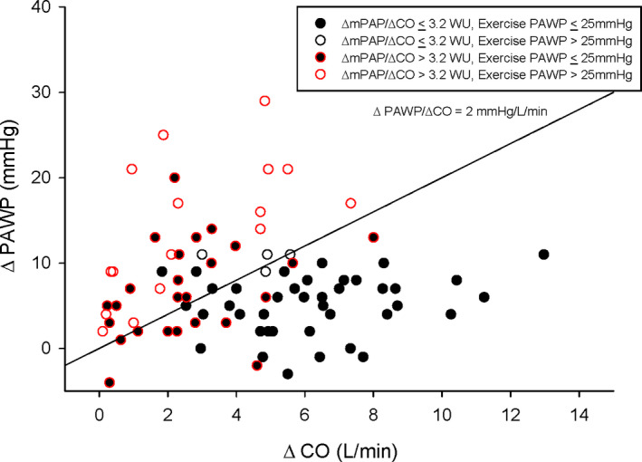
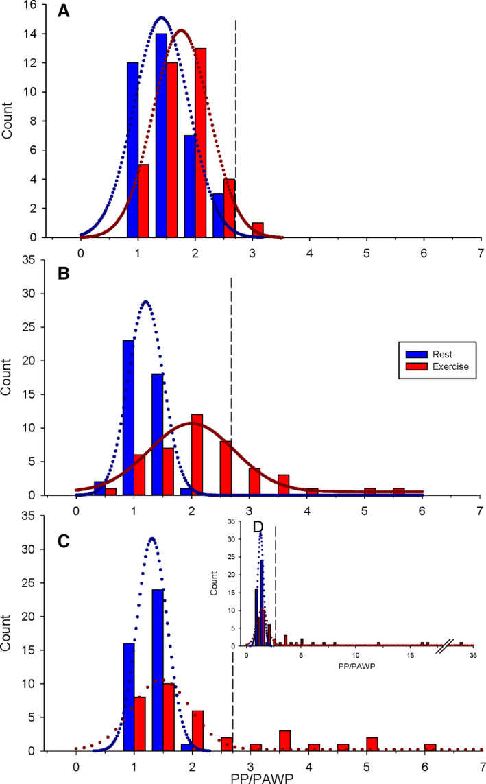
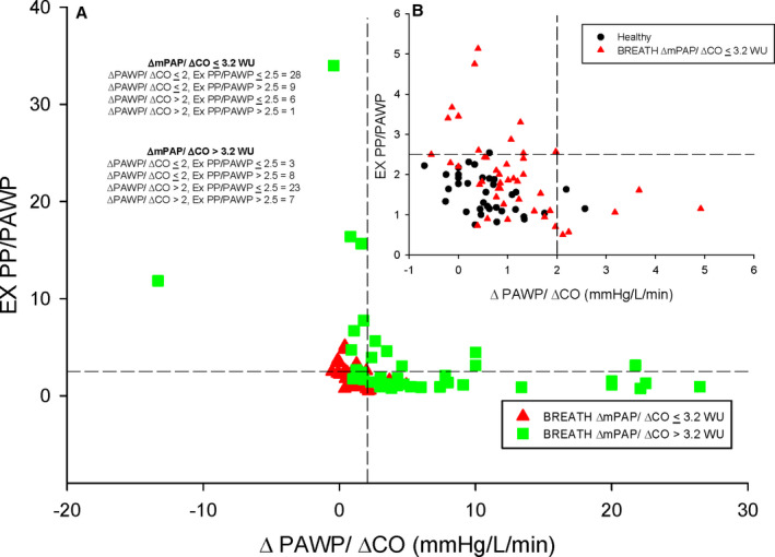
Background Resting right heart catheterization can assess both left heart filling and pulmonary artery (PA) pressures to identify and classify pulmonary hypertension. Although exercise may further elucidate hemodynamic abnormalities, current pulmonary hypertension classifications do not consider the expected interrelationship between PA and left heart filling pressures. This study explored the utility of this relationship to enhance the classification of exercise hemodynamic phenotypes in pulmonary hypertension. Methods and Results Data from 36 healthy individuals (55, 50-60 years, 50% male) and 85 consecutive patients (60, 49-71 years, 48% male) with dyspnea and/or suspected pulmonary hypertension of uncertain etiology were analyzed. Right heart catheterization was performed at rest and during semiupright submaximal cycling. To classify exercise phenotypes in patients, upper 95% CIs were identified from the healthy individuals for the change from rest to exercise in mean PA pressure over cardiac output (ΔmPAP/ΔCO ≤3.2 Wood units [WU]), pulmonary artery wedge pressure over CO (ΔPAWP/ΔCO ≤2 mm Hg/L per minute), and exercise PA pulse pressure over PAWP (PP/PAWP ≤2.5). Among patients with a ΔmPAP/ΔCO ≤3.2 WU, the majority (84%) demonstrated a ΔPAWP/ΔCO ≤2 mm Hg/L per minute, yet 23% demonstrated an exercise PP/PAWP >2.5. Among patients with a ΔmPAP/ΔCO >3.2 WU, 37% had an exercise PP/PAWP >2.5 split between ΔPAWP/ΔCO groups. Patients with normal hemodynamic classification declined from 52% at rest to 36% with exercise. Conclusions The addition of PP/PAWP to classify exercise hemodynamics uncovers previously unrecognized abnormal phenotypes within each ΔmPAP/ΔCO group. Our study refines abnormal exercise hemodynamic phenotypes based on an understanding of the interrelationship between PA and left heart filling pressures.
背景 静息右心导管检查可评估左心充盈压和肺动脉(PA)压,以识别和分类肺动脉高压。尽管运动可能进一步阐明血液动力学异常,但目前的肺动脉高压分类并不考虑 PA 和左心充盈压之间的预期相互关系。本研究探讨了这种关系在增强肺动脉高压运动血液动力学表型分类中的作用。
方法和结果 分析了 36 名健康个体(55 岁,50-60 岁,50%为男性)和 85 例连续呼吸困难和/或疑似肺动脉高压病因不明的患者的数据。在休息和半直立亚最大循环期间进行右心导管检查。为了对患者进行运动表型分类,从健康个体中确定了上 95%CI,用于评估从休息到运动时平均 PA 压与心输出量的变化(ΔmPAP/ΔCO≤3.2 伍德单位[WU])、PA 楔压与 CO 的变化(ΔPAWP/ΔCO≤2mmHg/L/分钟)以及运动时 PA 脉搏压与 PAWP 的变化(PP/PAWP≤2.5)。在ΔmPAP/ΔCO≤3.2 WU 的患者中,大多数(84%)表现为ΔPAWP/ΔCO≤2mmHg/L/分钟,但 23%表现为运动时 PP/PAWP>2.5。在ΔmPAP/ΔCO>3.2 WU 的患者中,37%的运动时 PP/PAWP>2.5,分为ΔPAWP/ΔCO 两组。正常血液动力学分类的患者从休息时的 52%下降到运动时的 36%。
结论 将 PP/PAWP 用于运动血液动力学分类可以揭示每个ΔmPAP/ΔCO 组中以前未被识别的异常表型。我们的研究基于对 PA 和左心充盈压之间相互关系的理解,对异常运动血液动力学表型进行了细化。