• 文献检索
  • 文档翻译
  • 深度研究
  • 学术资讯
  • Suppr Zotero 插件Zotero 插件
  • 邀请有礼
  • 套餐&价格
  • 历史记录
应用&插件
Suppr Zotero 插件Zotero 插件浏览器插件Mac 客户端Windows 客户端微信小程序
定价
高级版会员购买积分包购买API积分包
服务
文献检索文档翻译深度研究API 文档MCP 服务
关于我们
关于 Suppr公司介绍联系我们用户协议隐私条款
关注我们

Suppr 超能文献

核心技术专利:CN118964589B侵权必究
粤ICP备2023148730 号-1Suppr @ 2026

文献检索

告别复杂PubMed语法,用中文像聊天一样搜索,搜遍4000万医学文献。AI智能推荐,让科研检索更轻松。

立即免费搜索

文件翻译

保留排版,准确专业,支持PDF/Word/PPT等文件格式,支持 12+语言互译。

免费翻译文档

深度研究

AI帮你快速写综述,25分钟生成高质量综述,智能提取关键信息,辅助科研写作。

立即免费体验

正常眼压性青光眼中小动脉、小静脉和毛细血管变化的全球评估。

Global assessment of arteriolar, venular and capillary changes in normal tension glaucoma.

机构信息

Department of Ophthalmology and Visual Sciences, The Chinese University of Hong Kong, Hong Kong, China.

Hong Kong Eye Hospital, Hong Kong, China.

出版信息

Sci Rep. 2020 Nov 5;10(1):19222. doi: 10.1038/s41598-020-75784-1.

DOI:10.1038/s41598-020-75784-1
PMID:33154407
原文链接:https://pmc.ncbi.nlm.nih.gov/articles/PMC7644687/
Abstract

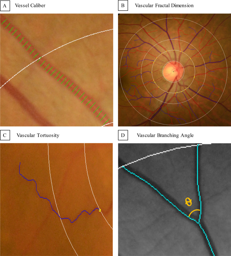
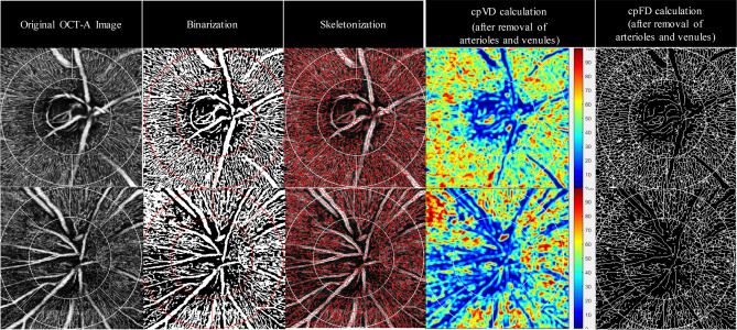
Microcirculatory insufficiency has been hypothesized in glaucoma pathogenesis. There is a scarcity of data to comprehensively examine the changes in retinal microvasculature and its role in normal tension glaucoma (NTG). We conducted a cross-sectional case-control study and included 168 eyes from 100 NTG patients and 68 healthy subjects. Quantitative retinal arteriolar and venular metrics were measured from retinal photographs using a computer-assisted program. Radial peripapillary capillary network was imaged with OCT-A and quantitative capillary metrics (circumpapillary vessel density (cpVD) and circumpapillary fractal dimension (cpFD)) were measured with a customized MATLAB program. We found that NTG was associated with decreased arteriolar and venular tortuosity, arteriolar branching angle, cpVD and cpFD. Decreased venular caliber, arteriolar and venular branching angles, cpVD and cpFD were associated with thinner average RNFL thickness. Decreased arteriolar and venular branching angles, cpVD and cpFD were also associated with worse standard automated perimetry measurements (mean deviation and visual field index). Compared with retinal arteriolar and venular metrics, regression models based on OCT-A capillary metrics consistently showed stronger associations with NTG and structural and functional measurements in NTG. We concluded that NTG eyes showed generalized microvascular attenuations, in which OCT-A capillary metrics attenuations were more prominent and strongly associated with NTG.

摘要

微循环不足在青光眼发病机制中被假设存在。目前,全面检查视网膜微血管变化及其在正常眼压性青光眼(NTG)中的作用的数据很少。我们进行了一项横断面病例对照研究,纳入了 100 名 NTG 患者和 68 名健康对照者的 168 只眼。使用计算机辅助程序从视网膜照片中测量定量视网膜动脉和静脉血管参数。使用 OCT-A 对视盘周围毛细血管网络进行成像,并使用定制的 MATLAB 程序测量毛细血管定量参数(环视盘血管密度(cpVD)和环视盘分形维数(cpFD))。我们发现,NTG 与视网膜小动脉和小静脉迂曲度、小动脉分支角、cpVD 和 cpFD 降低有关。小静脉口径、小动脉和小静脉分支角、cpVD 和 cpFD 的降低与平均 RNFL 厚度变薄有关。小动脉和小静脉分支角、cpVD 和 cpFD 的降低也与标准自动视野计测量值(平均偏差和视野指数)更差有关。与视网膜小动脉和小静脉参数相比,基于 OCT-A 毛细血管参数的回归模型与 NTG 以及 NTG 中的结构和功能测量值具有更强的相关性。我们得出结论,NTG 眼表现出广泛的微血管衰减,其中 OCT-A 毛细血管参数衰减更为明显且与 NTG 强烈相关。

https://cdn.ncbi.nlm.nih.gov/pmc/blobs/fff7/7644687/48c267b74262/41598_2020_75784_Fig2_HTML.jpg
https://cdn.ncbi.nlm.nih.gov/pmc/blobs/fff7/7644687/823f5f432e9a/41598_2020_75784_Fig1_HTML.jpg
https://cdn.ncbi.nlm.nih.gov/pmc/blobs/fff7/7644687/48c267b74262/41598_2020_75784_Fig2_HTML.jpg
https://cdn.ncbi.nlm.nih.gov/pmc/blobs/fff7/7644687/823f5f432e9a/41598_2020_75784_Fig1_HTML.jpg
https://cdn.ncbi.nlm.nih.gov/pmc/blobs/fff7/7644687/48c267b74262/41598_2020_75784_Fig2_HTML.jpg

相似文献

1
Global assessment of arteriolar, venular and capillary changes in normal tension glaucoma.正常眼压性青光眼中小动脉、小静脉和毛细血管变化的全球评估。
Sci Rep. 2020 Nov 5;10(1):19222. doi: 10.1038/s41598-020-75784-1.
2
Comparison of optical coherence tomography angiography metrics in primary angle-closure glaucoma and normal-tension glaucoma.原发性闭角型青光眼和正常眼压型青光眼的光学相干断层扫描血管造影指标比较。
Sci Rep. 2021 Nov 30;11(1):23136. doi: 10.1038/s41598-021-02296-x.
3
The association between retinal vessel diameter and retinal nerve fiber layer thickness in asymmetric normal tension glaucoma patients.双眼不对称正常眼压性青光眼患者视网膜血管直径与视网膜神经纤维层厚度的相关性。
Invest Ophthalmol Vis Sci. 2012 Aug 17;53(9):5609-14. doi: 10.1167/iovs.12-9783.
4
Comparison of retinal microvascular changes in eyes with high-tension glaucoma or normal-tension glaucoma: a quantitative optic coherence tomography angiographic study.高眼压性青光眼或正常眼压性青光眼患者眼部视网膜微血管变化的比较:一项定量光学相干断层扫描血管造影研究
Graefes Arch Clin Exp Ophthalmol. 2018 Jun;256(6):1179-1186. doi: 10.1007/s00417-018-3930-z. Epub 2018 Feb 15.
5
Microvascular Compromise Develops Following Nerve Fiber Layer Damage in Normal-Tension Glaucoma Without Choroidal Vasculature Involvement.在无脉络膜血管受累的正常眼压青光眼中,神经纤维层损伤后会发生微血管损害。
J Glaucoma. 2017 Mar;26(3):216-222. doi: 10.1097/IJG.0000000000000587.
6
Relationship between Optical Coherence Tomography Angiography Vessel Density and Severity of Visual Field Loss in Glaucoma.光学相干断层扫描血管造影血管密度与青光眼视野缺损严重程度之间的关系
Ophthalmology. 2016 Dec;123(12):2498-2508. doi: 10.1016/j.ophtha.2016.08.041. Epub 2016 Oct 7.
7
Global Assessment of Retinal Arteriolar, Venular and Capillary Microcirculations Using Fundus Photographs and Optical Coherence Tomography Angiography in Diabetic Retinopathy.利用眼底照相和光相干断层扫描血管造影术评估糖尿病视网膜病变的视网膜小动脉、小静脉和毛细血管微循环。
Sci Rep. 2019 Aug 13;9(1):11751. doi: 10.1038/s41598-019-47770-9.
8
Peripapillary and Macular Vessel Density Measurement by Optical Coherence Tomography Angiography in Pseudoexfoliation and Primary Open-angle Glaucoma.光学相干断层扫描血管造影术在假性剥脱综合征和原发性开角型青光眼的周边和黄斑血管密度测量。
J Glaucoma. 2020 May;29(5):381-385. doi: 10.1097/IJG.0000000000001464.
9
Optical Coherence Tomography Angiography Analysis of Perfused Peripapillary Capillaries in Primary Open-Angle Glaucoma and Normal-Tension Glaucoma.原发性开角型青光眼和正常眼压性青光眼视乳头周围灌注毛细血管的光学相干断层扫描血管造影分析
Invest Ophthalmol Vis Sci. 2016 Jul 1;57(9):OCT611-OCT620. doi: 10.1167/iovs.15-18945.
10
Risk of Normal Tension Glaucoma Progression From Automated Baseline Retinal-Vessel Caliber Analysis: A Prospective Cohort Study.基于自动基线视网膜血管管径分析的正常眼压性青光眼进展风险:一项前瞻性队列研究。
Am J Ophthalmol. 2023 Mar;247:111-120. doi: 10.1016/j.ajo.2022.09.015. Epub 2022 Oct 9.

引用本文的文献

1
Computerized Analysis of the Eye Vasculature in a Mass Dataset of Digital Fundus Images: The Example of Age, Sex, and Primary Open-Angle Glaucoma.数字化眼底图像大规模数据集中眼部血管系统的计算机分析:以年龄、性别和原发性开角型青光眼为例
Ophthalmol Sci. 2025 Mar 28;5(5):100778. doi: 10.1016/j.xops.2025.100778. eCollection 2025 Sep-Oct.
2
Repeatability, interocular correlation and agreement of optic nerve head vessel density in healthy eyes: a swept-source optical coherence tomographic angiography study.健康眼睛视神经乳头血管密度的可重复性、眼间相关性及一致性:一项扫频源光学相干断层血管造影研究
Int J Ophthalmol. 2024 May 18;17(5):896-903. doi: 10.18240/ijo.2024.05.14. eCollection 2024.
3

本文引用的文献

1
Latest Developments in Normal-Pressure Glaucoma: Diagnosis, Epidemiology, Genetics, Etiology, Causes and Mechanisms to Management.正常眼压性青光眼的最新进展:诊断、流行病学、遗传学、病因学、发病机制和治疗。
Asia Pac J Ophthalmol (Phila). 2019 Nov-Dec;8(6):457-468. doi: 10.1097/01.APO.0000605096.48529.9c.
2
Global Blindness: The Progress We Are Making and Still Need to Make.全球失明状况:我们已经取得的进展和仍需做出的努力。
Asia Pac J Ophthalmol (Phila). 2019 Nov-Dec;8(6):424-428. doi: 10.1097/APO.0000000000000264.
3
Normal Pressure Glaucoma: The Challenge in Asia and the Scientific Contributions from Asia.
Is Primary Open-Angle Glaucoma a Vascular Disease? Assessment of the Relationship between Retinal Arteriolar Morphology and Glaucoma Severity Using Adaptive Optics.
原发性开角型青光眼是一种血管疾病吗?利用自适应光学评估视网膜小动脉形态与青光眼严重程度之间的关系。
J Clin Med. 2024 Jan 15;13(2):478. doi: 10.3390/jcm13020478.
4
Quantification of vascular morphology in optical coherence tomography angiography in primary open angle glaucoma.原发性开角型青光眼光学相干断层扫描血管造影中血管形态的量化分析
Adv Ophthalmol Pract Res. 2023 May 12;3(3):119-125. doi: 10.1016/j.aopr.2023.05.002. eCollection 2023 Aug-Sep.
5
The Differences in the Pattern of OCT and OCTA Examinations between Early Normal- and High-Tension Pseudoexfoliative Glaucoma.早期正常眼压性和高眼压性假性剥脱性青光眼在光学相干断层扫描(OCT)和光学相干断层扫描血管造影(OCTA)检查模式上的差异
J Clin Med. 2023 Jul 26;12(15):4899. doi: 10.3390/jcm12154899.
6
Deep Learning in Optical Coherence Tomography Angiography: Current Progress, Challenges, and Future Directions.光学相干断层扫描血管造影中的深度学习:当前进展、挑战及未来方向。
Diagnostics (Basel). 2023 Jan 16;13(2):326. doi: 10.3390/diagnostics13020326.
7
Ocular Factors of Fractal Dimension and Blood Vessel Tortuosity Derived From OCTA in a Healthy Chinese Population.基于 OCTA 的中国人眼分形维数和血管迂曲度的眼部影响因素。
Transl Vis Sci Technol. 2022 May 2;11(5):1. doi: 10.1167/tvst.11.5.1.
正常眼压性青光眼:亚洲面临的挑战及亚洲的科学贡献
Asia Pac J Ophthalmol (Phila). 2019 Nov-Dec;8(6):419-421. doi: 10.1097/01.APO.0000605104.33282.14.
4
Optical Coherence Tomography for the Diagnosis and Monitoring of Glaucoma.用于青光眼诊断和监测的光学相干断层扫描
Asia Pac J Ophthalmol (Phila). 2019;8(2):135-145. doi: 10.22608/APO.201902.
5
Optical Coherence Tomography Angiography and Glaucoma: A Brief Review.光学相干断层扫描血管造影与青光眼:简要综述
Asia Pac J Ophthalmol (Phila). 2019;8(2):115-125. doi: 10.22608/APO.201914.
6
Optical Coherence Tomography Angiography Compared With Optical Coherence Tomography Macular Measurements for Detection of Glaucoma.光学相干断层扫描血管造影与光学相干断层扫描黄斑测量在青光眼检测中的比较。
JAMA Ophthalmol. 2018 Aug 1;136(8):866-874. doi: 10.1001/jamaophthalmol.2018.1627.
7
Foveal Avascular Zone in Normal Tension Glaucoma Measured by Optical Coherence Tomography Angiography.光学相干断层扫描血管造影测量正常眼压性青光眼的中心凹无血管区。
Biomed Res Int. 2017;2017:3079141. doi: 10.1155/2017/3079141. Epub 2017 Dec 17.
8
Comparison of peripapillary vessel density between preperimetric and perimetric glaucoma evaluated by OCT-angiography.通过光学相干断层扫描血管造影术评估的视野检查前和视野检查时青光眼患者视乳头周围血管密度的比较。
PLoS One. 2017 Aug 31;12(8):e0184297. doi: 10.1371/journal.pone.0184297. eCollection 2017.
9
Peripapillary and Macular Vessel Density in Patients with Glaucoma and Single-Hemifield Visual Field Defect.青光眼和单半视野视觉缺损患者的视乳头周围和黄斑区血管密度
Ophthalmology. 2017 May;124(5):709-719. doi: 10.1016/j.ophtha.2017.01.004. Epub 2017 Feb 10.
10
Microvascular Compromise Develops Following Nerve Fiber Layer Damage in Normal-Tension Glaucoma Without Choroidal Vasculature Involvement.在无脉络膜血管受累的正常眼压青光眼中,神经纤维层损伤后会发生微血管损害。
J Glaucoma. 2017 Mar;26(3):216-222. doi: 10.1097/IJG.0000000000000587.