Department of Immunobiology, Institute of Biology Sciences, Maria Curie-Skłodowska University, Akademicka 19, 20-033, Lublin, Poland.
Department of Earth Sciences, Graduate School of Science, Chiba University, Chiba, Japan.
Sci Rep. 2020 Nov 5;10(1):19167. doi: 10.1038/s41598-020-76215-x.
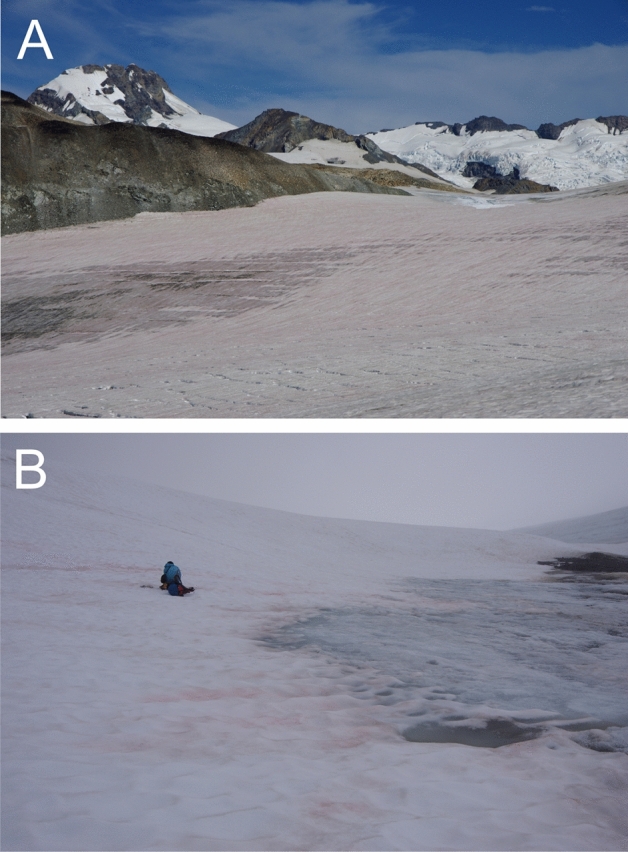
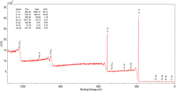
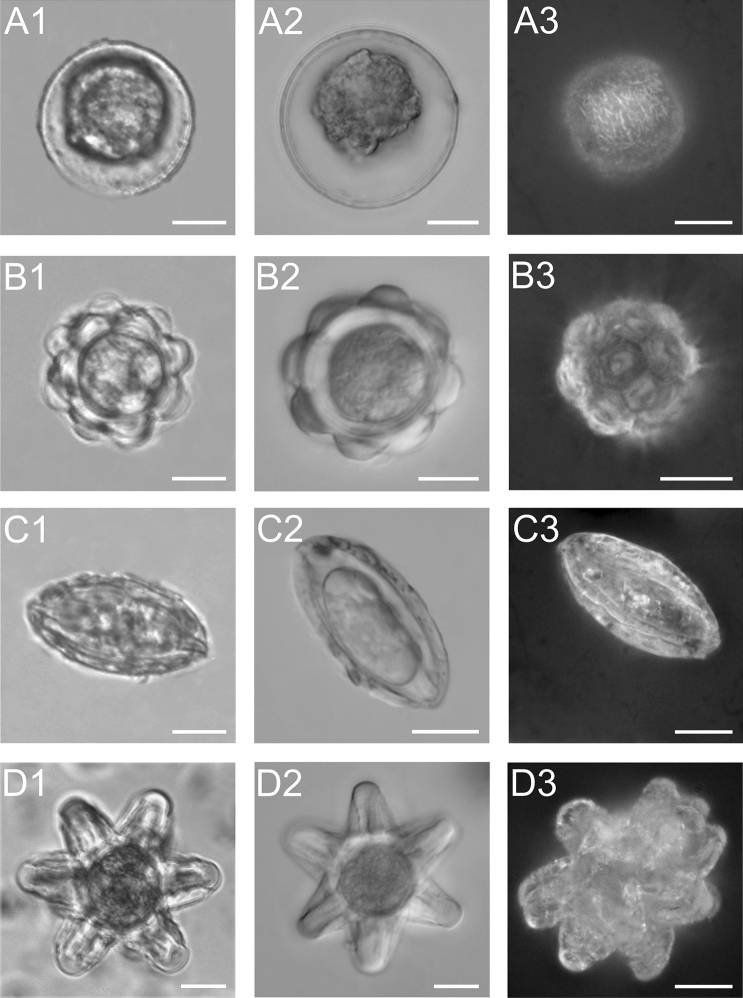
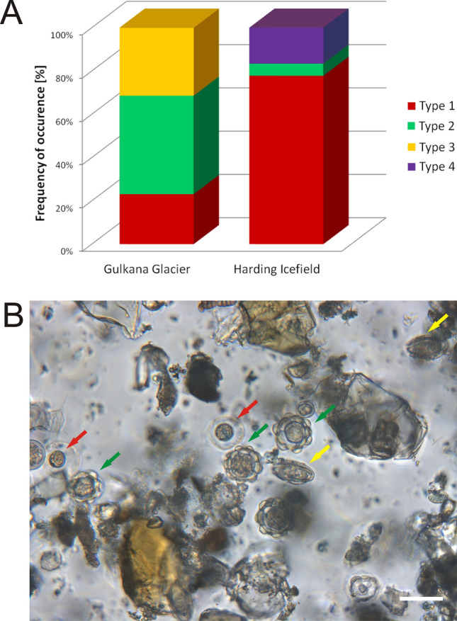
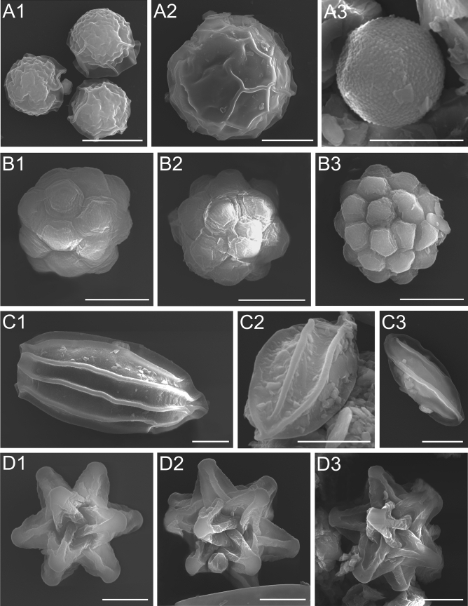
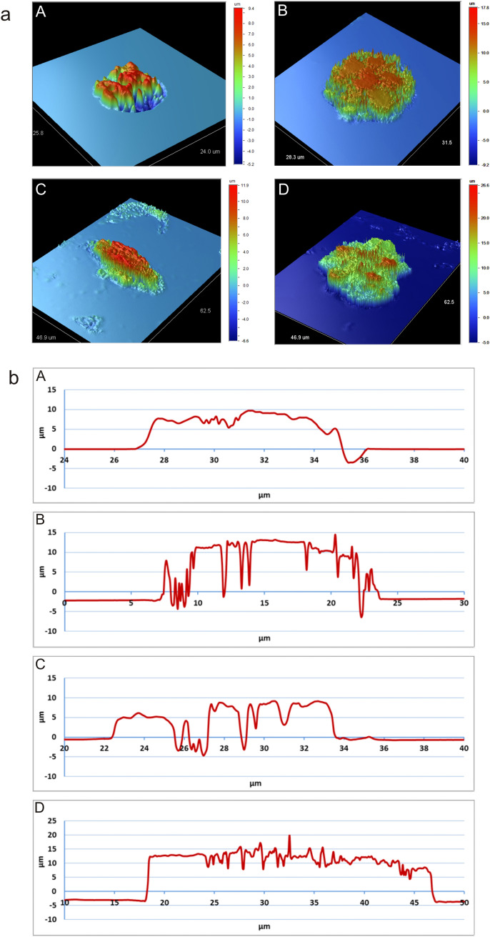
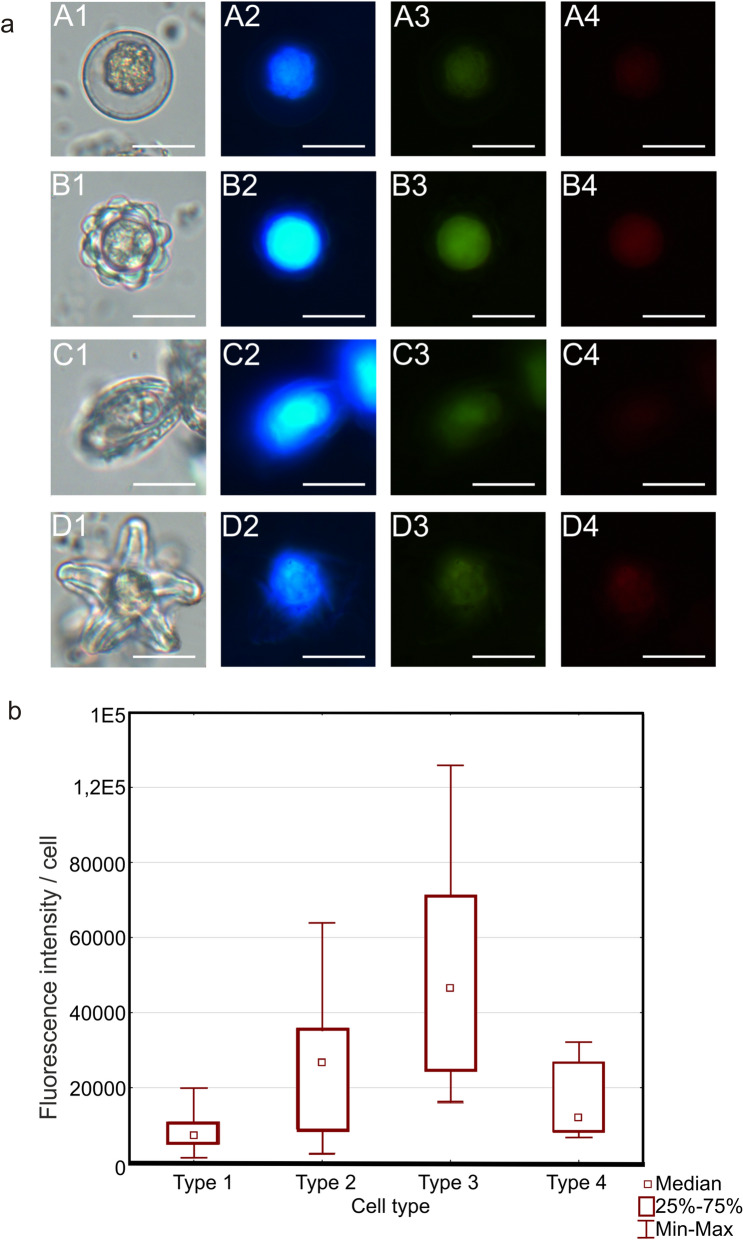
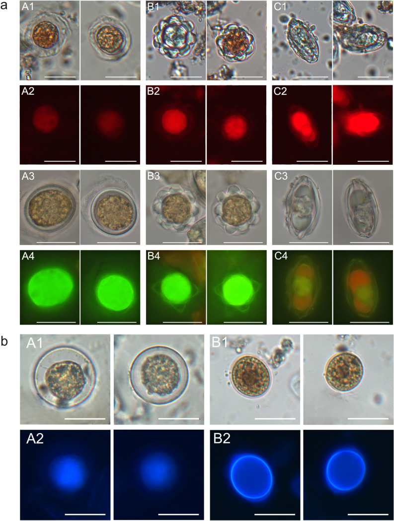
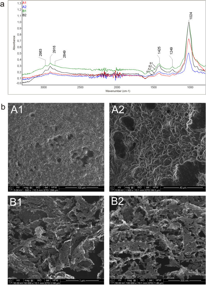
Snow algae are photosynthetic microbes growing in thawing snow. They usually show various morphological cell types. The aim of this study was to carry out microscopic and spectroscopic analysis of different forms of cells of snow algae collected on glaciers in Alaska. Four different shapes of algal cells were observed with the use of bright field LM (Light Microscopy), DIC (Differential Interference Contrast), EDF (Extended Depth Focus), fluorescence microscopy, and SEM (Scanning Electron Microscopy). The cells exhibited the strongest autofluorescence after the exposure to 365-nm excitation light, and the intensity differed among the cell types. Zygotes (cysts) showed the most intense fluorescence. Acridine orange staining revealed the acid nature of the algal cells. The use of Congo red and Calcofluor white fluorochromes indicated differences in the structure of polysaccharides in the cell wall in the individual types of algal cells. FTIR (Fourier-Transform Infrared Spectroscopy) analyses showed the presence of polysaccharides not only in the algal cells but also in the fixative solution. The presence of polysaccharides in the extracellular algal fraction was confirmed by X-ray dispersion spectroscopy (EDS), X-ray photoelectron spectroscopy (XPS), and scanning electron microscopy imaging (SEM). The differences observed in the structure of the cell wall of the different forms of red snow algae prompt further analysis of this structure.
雪藻是在融雪中生长的光合微生物。它们通常表现出各种形态的细胞类型。本研究的目的是对在阿拉斯加冰川上采集的雪藻的不同细胞形式进行显微镜和光谱分析。使用明场 LM(光学显微镜)、DIC(微分干涉对比)、EDF(扩展景深)、荧光显微镜和 SEM(扫描电子显微镜)观察到四种不同形状的藻类细胞。细胞在暴露于 365nm 激发光后表现出最强的自发荧光,并且不同细胞类型之间的强度不同。受精卵(休眠孢)显示出最强烈的荧光。吖啶橙染色显示藻类细胞呈酸性。刚果红和 Calcofluor 白荧光染料的使用表明,不同类型的藻类细胞的细胞壁多糖结构存在差异。FTIR(傅里叶变换红外光谱)分析表明,不仅在藻类细胞中,而且在固定剂溶液中都存在多糖。X 射线漫散射光谱(EDS)、X 射线光电子能谱(XPS)和扫描电子显微镜成像(SEM)证实了细胞外藻类部分存在多糖。不同形式的红色雪藻细胞壁结构的差异促使对这种结构进行进一步分析。