Interdisciplinary Center for Gene Therapy, Escola Paulista de Medicina, Universidade Federal de São Paulo, São Paulo, Brazil.
Department of Physiological Sciences, Faculdade de Ciências Medicas da Santa Casa de São Paulo, São Paulo, Brazil.
Stem Cell Res Ther. 2020 Nov 6;11(1):473. doi: 10.1186/s13287-020-01992-1.
After traumatic skeletal muscle injury, muscle healing is often incomplete and produces extensive fibrosis. The sequence of M1 and M2 macrophage accumulation and the duration of each subtype in the injured area may help to direct the relative extent of fibrogenesis and myogenesis during healing. We hypothesized that increasing the number of M1 macrophages early after traumatic muscle injury would produce more cellular and molecular substrates for myogenesis and fewer substrates for fibrosis, leading to better muscle healing.
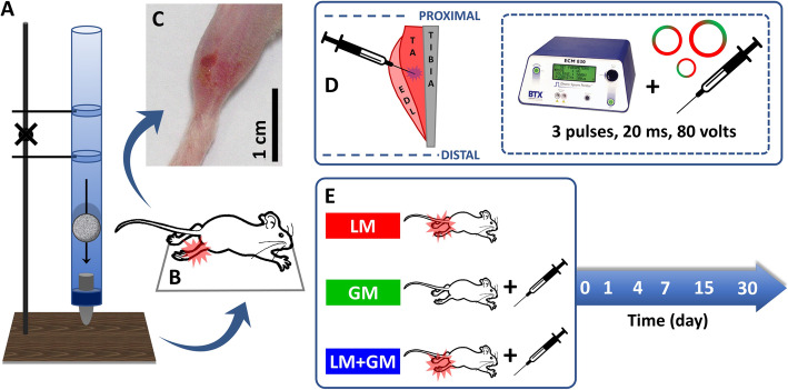
To test this hypothesis, we transfected skeletal muscle with a plasmid vector to transiently express GM-CSF shortly after injury to drive the polarization of macrophages towards the M1 subset. C57BL/6 mouse tibialis anterior (TA) muscles were injured by contusion and electroporated with uP-mGM, which is a plasmid vector that transiently expresses GM-CSF. Myogenesis, angiogenesis, and fibrosis were evaluated by histology, immunohistochemistry, and RT-qPCR; subpopulations of macrophages by flow cytometry; and muscle functioning by the maximum running speed on the treadmill and the recovery of muscle mass.
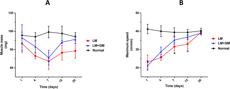
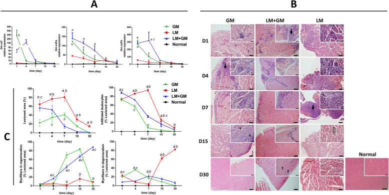
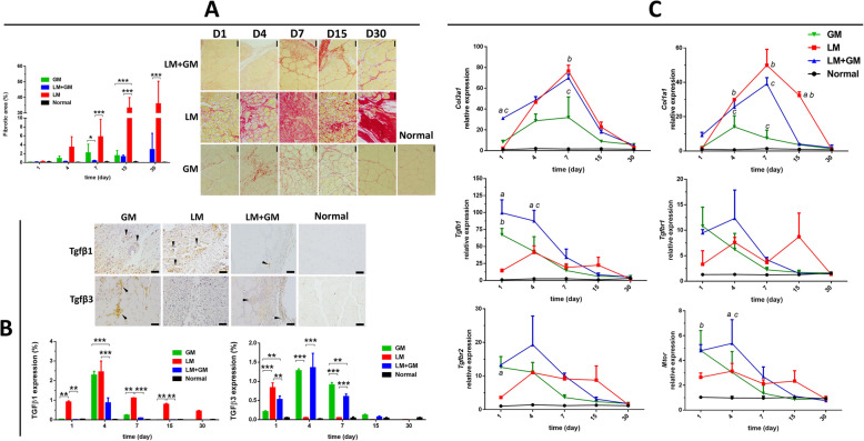
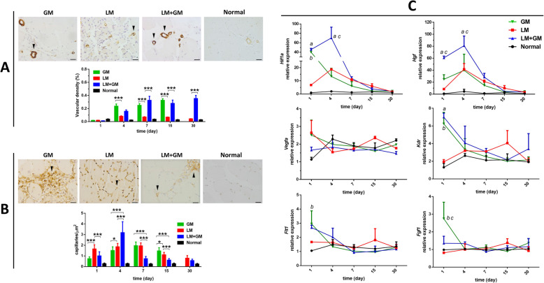
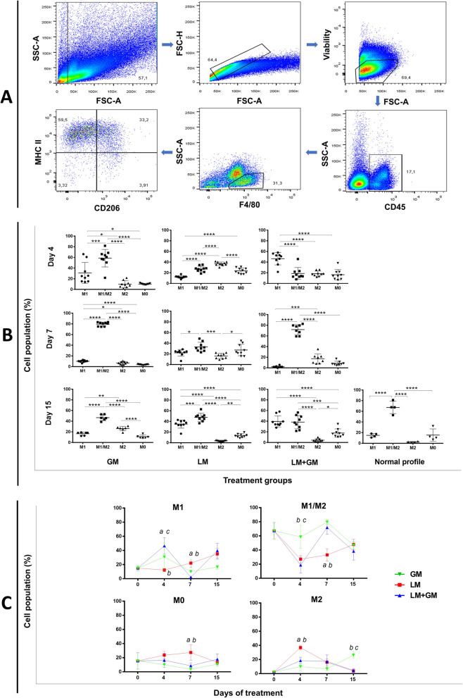
Muscle injury increased the number of local M1-like macrophages and decreased the number of M2-like macrophages on day 4, and uP-mGM treatment enhanced this variation. uP-mGM treatment decreased TGF-β1 protein expression on day 4, and the Sirius Red-positive area decreased from 35.93 ± 15.45% (no treatment) to 2.9% ± 6.5% (p < 0.01) on day 30. uP-mGM electroporation also increased Hgf, Hif1α, and Mtor gene expression; arteriole density; and muscle fiber number during regeneration. The improvement in the quality of the muscle tissue after treatment with uP-mGM affected the increase in the TA muscle mass and the maximum running speed on a treadmill.
Collectively, our data show that increasing the number of M1-like macrophages immediately after traumatic muscle injury promotes muscle recovery with less fibrosis, and this can be achieved by the transient expression of GM-CSF.
在创伤性骨骼肌损伤后,肌肉愈合通常不完整,并产生广泛的纤维化。在受伤区域中 M1 和 M2 巨噬细胞积累的顺序和每种亚型的持续时间可能有助于指导愈合过程中纤维化和肌生成的相对程度。我们假设在创伤性肌肉损伤后早期增加 M1 巨噬细胞的数量将为肌生成产生更多的细胞和分子底物,为纤维化产生更少的底物,从而导致更好的肌肉愈合。
为了验证这一假设,我们在损伤后不久通过转染骨骼肌的质粒载体瞬时表达 GM-CSF,以驱动巨噬细胞向 M1 亚群极化。C57BL/6 小鼠的胫骨前肌(TA)通过挫伤和电穿孔用 uP-mGM 损伤,uP-mGM 是一种瞬时表达 GM-CSF 的质粒载体。通过组织学、免疫组织化学和 RT-qPCR 评估肌生成、血管生成和纤维化;通过流式细胞术评估巨噬细胞亚群;通过跑步机上的最大跑步速度和肌肉质量的恢复评估肌肉功能。
肌肉损伤导致第 4 天局部 M1 样巨噬细胞数量增加,M2 样巨噬细胞数量减少,uP-mGM 处理增强了这种变化。uP-mGM 处理降低了第 4 天的 TGF-β1 蛋白表达,天狼星红阳性面积从 35.93±15.45%(无处理)减少到 30 天的 2.9%±6.5%(p<0.01)。uP-mGM 电穿孔还增加了 Hgf、Hif1α 和 Mtor 基因表达、小动脉密度和再生过程中的肌纤维数量。uP-mGM 处理后肌肉组织质量的改善影响了 TA 肌肉质量和跑步机上最大跑步速度的增加。
总之,我们的数据表明,在创伤性肌肉损伤后立即增加 M1 样巨噬细胞的数量可促进肌肉恢复,纤维化程度降低,这可以通过 GM-CSF 的瞬时表达来实现。