Department of Ophthalmology, Cheng Hsin General Hospital, Taipei, Taiwan.
Institute of Pharmacology, School of Medicine, National Yang-Ming University, Taipei, Taiwan.
BMC Complement Med Ther. 2020 Nov 10;20(1):338. doi: 10.1186/s12906-020-03136-7.
Emodin has been proved to have an anti-ischemic effect on the brain, however little research has been done on its effect on vision-threatening retinal ischemia. Thus, an investigation was carried out into the hypothetical efficacy of emodin against retinal ischemia and the role of β-catenin/VEGF in its therapeutic mechanism.
Retinal ischemia, followed by reperfusion (IR), was inducted by raising the intraocular pressure of a Wistar rat's eye to 120 mmHg for 60 min. Additionally, pre-ischemic/post-ischemic intravitreous injections of emodin (4, 10 and 20 μM) or vehicle were carried out on the eye with retinal ischemia. MTT assay, electroretinograms, cresyl violet staining retinal thickness measurements, and fluorogold retrograde labelling of retinal ganglion cells (RGCs) as well as Western blotting were carried out.
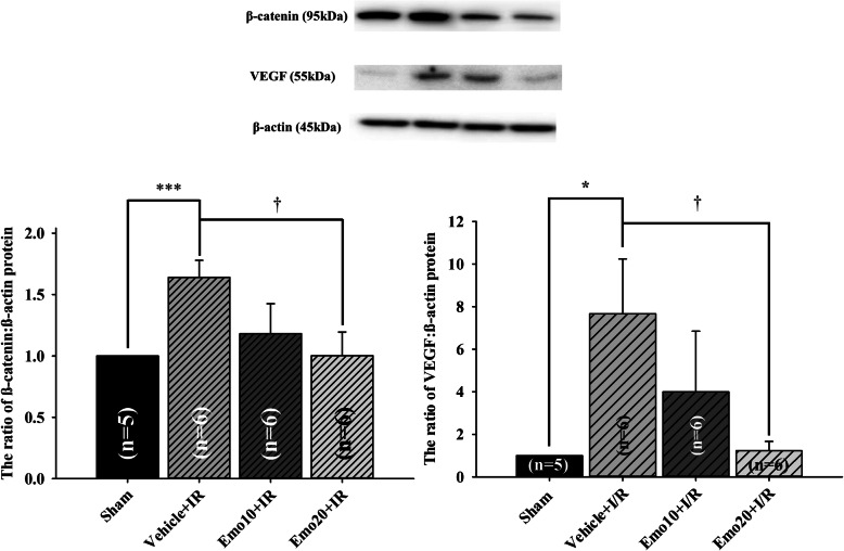
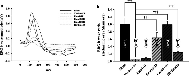
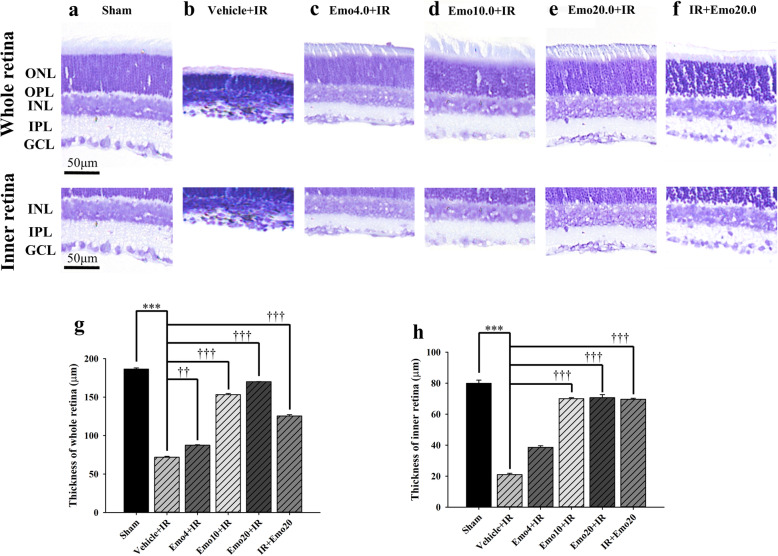
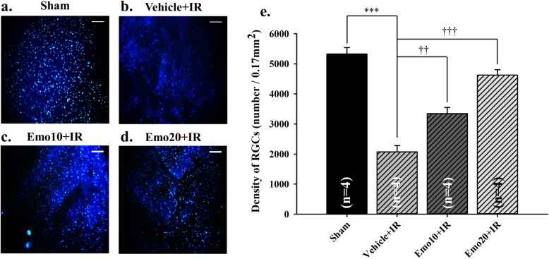
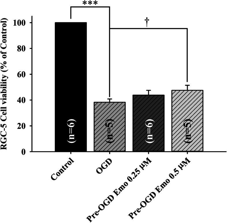
Cultured RGC-5 cells subjected to oxygen glucose deprivation (OGD) were used to confirm the effective concentrations of emodin (administered 1 h pre-OGD, pre-OGD emodin). The most effective and significant (P = 0.04) dose of pre-OGD emodin was observed at 0.5 μM (cell viability: 47.52 ± 3.99%) as compared to pre-OGD vehicle treatment group (38.30 ± 2.51%). Furthermore, pre-ischemic intravitreous injection of 20 μM emodin (Emo20 + IR = 0.99 ± 0.18, P < 0.001) significantly attenuated the ischemia induced reduction in ERG b-wave amplitude, as compared to pre-ischemic intravitreous vehicle (Vehicle+IR = 0.04 ± 0.02). Post-ischemic intravitreous 20 μM emodin also significantly (P < 0.001) attenuated the ischemia associated b-wave reduction (IR + Em20 = 0.24 ± 0.09). Compared with pre-ischemic intravitreous vehicle (Vehicle+IR; whole retina thickness = 71.80 ± 1.08 μm; inner retina thickness = 20.97 ± 0.85 μm; RGC =2069.12 ± 212.82/0.17mm), the significant (P < 0.001) protective effect was also present with pre-ischemic administration of emodin. This was shown by observing cresyl violet stained retinal thickness (Emo20 + IR: whole retina = 170.10 ± 0.10 μm; inner retina = 70.65 ± 2.06 μm) and retrograde fluorogold immunolabeled RGC density (4623.53 ± 179.48/0.17mm). As compared to the normal control (the ratio of β-catenin/VEGF to β-actin was set as 1 in the Sham group), the β-catenin/VEGF protein level significantly (P < 0.001) increased after retinal ischemia and when pre-ischemic intravitreous vehicle (Vehicle+IR = 1.64 ± 0.14/7.67 ± 2.57) was carried out. However, these elevations were significantly (P = 0.02) attenuated by treatment with emodin 20 μM (Emo20 + IR = 1.00 ± 0.19/1.23 ± 0.44).
The present results suggest that emodin might protect against retinal ischemia insulted neurons such as RGCs by significantly downregulating the upregulation of β-catenin/VEGF protein that occurs during ischemia.
已经证明大黄素对大脑具有抗缺血作用,但对威胁视力的视网膜缺血的作用研究甚少。因此,本研究旨在调查大黄素对视网膜缺血的假设疗效以及β-连环蛋白/VEGF 在其治疗机制中的作用。
通过将 Wistar 大鼠的眼内压升高至 120mmHg 持续 60 分钟来诱导视网膜缺血,再灌注(IR)。此外,在视网膜缺血的眼睛上进行预缺血/后缺血玻璃体腔内注射大黄素(4、10 和 20μM)或载体。进行 MTT 测定、视网膜电图、甲苯胺蓝染色视网膜厚度测量、视网膜神经节细胞(RGC)的荧光金逆行标记以及 Western blot 分析。
使用氧葡萄糖剥夺(OGD)处理的培养的 RGC-5 细胞来确认大黄素的有效浓度(在 OGD 前 1 小时给药,OGD 前大黄素)。观察到最有效和最显著(P=0.04)的预 OGD 大黄素剂量为 0.5μM(细胞活力:47.52±3.99%),与预 OGD 载体处理组(38.30±2.51%)相比。此外,在缺血前玻璃体腔内注射 20μM 大黄素(Emo20+IR=0.99±0.18,P<0.001)可显著减轻缺血引起的 ERG b 波幅度降低,与缺血前玻璃体腔内载体(Vehicle+IR=0.04±0.02)相比。后缺血玻璃体腔内 20μM 大黄素也显著(P<0.001)减轻了与缺血相关的 b 波降低(IR+Em20=0.24±0.09)。与缺血前玻璃体腔内载体(Vehicle+IR;整个视网膜厚度=71.80±1.08μm;内视网膜厚度=20.97±0.85μm;RGC=2069.12±212.82/0.17mm)相比,预缺血给予大黄素也具有显著(P<0.001)的保护作用。这通过观察甲苯胺蓝染色的视网膜厚度(Emo20+IR:整个视网膜=170.10±0.10μm;内视网膜=70.65±2.06μm)和逆行荧光金免疫标记的 RGC 密度(4623.53±179.48/0.17mm)得到证实。与正常对照组(Sham 组中β-连环蛋白/VEGF 与β-肌动蛋白的比值设为 1)相比,视网膜缺血后β-连环蛋白/VEGF 蛋白水平显著(P<0.001)升高,同时进行缺血前玻璃体腔内载体(Vehicle+IR=1.64±0.14/7.67±2.57)处理。然而,用 20μM 大黄素处理可显著(P=0.02)减弱这些升高(Emo20+IR=1.00±0.19/1.23±0.44)。
本研究结果表明,大黄素可能通过显著下调β-连环蛋白/VEGF 蛋白的上调,从而保护缺血性损伤的神经元,如 RGC,从而对视网膜缺血产生保护作用。