• 文献检索
  • 文档翻译
  • 深度研究
  • 学术资讯
  • Suppr Zotero 插件Zotero 插件
  • 邀请有礼
  • 套餐&价格
  • 历史记录
应用&插件
Suppr Zotero 插件Zotero 插件浏览器插件Mac 客户端Windows 客户端微信小程序
定价
高级版会员购买积分包购买API积分包
服务
文献检索文档翻译深度研究API 文档MCP 服务
关于我们
关于 Suppr公司介绍联系我们用户协议隐私条款
关注我们

Suppr 超能文献

核心技术专利:CN118964589B侵权必究
粤ICP备2023148730 号-1Suppr @ 2026

文献检索

告别复杂PubMed语法,用中文像聊天一样搜索,搜遍4000万医学文献。AI智能推荐,让科研检索更轻松。

立即免费搜索

文件翻译

保留排版,准确专业,支持PDF/Word/PPT等文件格式,支持 12+语言互译。

免费翻译文档

深度研究

AI帮你快速写综述,25分钟生成高质量综述,智能提取关键信息,辅助科研写作。

立即免费体验

基于生物发生的细胞外囊泡分类的定量蛋白质组学分析

Quantitative Proteomic Analysis of Biogenesis-Based Classification for Extracellular Vesicles.

作者信息

Zhang Linwen, Parot Jeremie, Hackley Vincent A, Turko Illarion V

机构信息

Biomolecular Measurement Division, National Institute of Standards and Technology, Gaithersburg, MD 20899, USA.

Institute for Bioscience and Biotechnology Research, Rockville, MD 20850, USA.

出版信息

Proteomes. 2020 Nov 6;8(4):33. doi: 10.3390/proteomes8040033.

DOI:10.3390/proteomes8040033
PMID:33171920
原文链接:https://pmc.ncbi.nlm.nih.gov/articles/PMC7709127/
Abstract

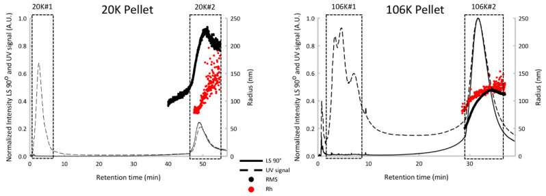
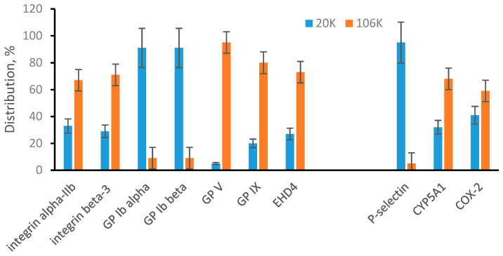
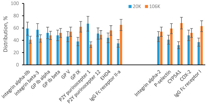
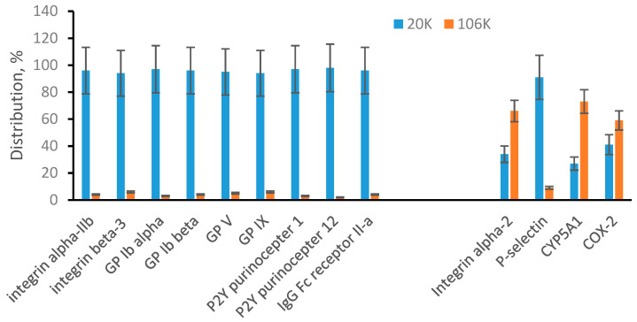
Extracellular vesicles (EVs) are traditionally divided into two major groups: (i) large vesicles originating from plasma membrane and called microvesicles, and (ii) small vesicles originating from the endoplasmic membrane and called exosomes. However, it is increasingly clear that the actual composition of a particular EV preparation cannot be adequately described with these two simple terms and is much more complex. Since the cell membrane origin of EVs predetermines their biological functions, the understanding of EV biogenesis is important for accurate interpretation of observed results. In the present study, we propose to take advantage of selective expression of some proteins in plasma or endosomal membranes and to use these proteins as plasma membrane-specific or endosomal membrane-specific markers. We have demonstrated that a quantitative mass spectrometry analysis allows simultaneous measurement of plasma membrane-specific and endosomal membrane-specific proteins in microvesicles and exosomes obtained after differential ultracentrifugation. Before mass spectrometry analysis, we also used sonicated platelets as a model of mixed EVs and multidetector asymmetrical-flow field-flow fractionation as an analytical method to verify a possible cross contamination of obtained microvesicles and exosomes. Based on the quantitative appearance of membrane-specific protein markers in EV preparations from human plasma and from human ARPE-19 cell medium, we concluded that there is no actual size limitation and both microvesicles and exosomes can be represented by large and small vesicles.

摘要

细胞外囊泡(EVs)传统上分为两大类:(i)源自质膜的大囊泡,称为微囊泡;(ii)源自内质膜的小囊泡,称为外泌体。然而,越来越明显的是,用这两个简单的术语无法充分描述特定EV制剂的实际组成,其要复杂得多。由于EVs的细胞膜起源决定了它们的生物学功能,因此了解EV的生物发生对于准确解释观察结果很重要。在本研究中,我们建议利用某些蛋白质在质膜或内体膜中的选择性表达,并将这些蛋白质用作质膜特异性或内体膜特异性标记物。我们已经证明,定量质谱分析可以同时测量差速超速离心后获得的微囊泡和外泌体中的质膜特异性和内体膜特异性蛋白质。在进行质谱分析之前,我们还使用超声处理的血小板作为混合EVs的模型,并使用多检测器不对称流场流分馏作为分析方法,以验证获得的微囊泡和外泌体是否可能存在交叉污染。基于来自人血浆和人ARPE-19细胞培养基的EV制剂中膜特异性蛋白质标记物的定量出现,我们得出结论,实际上不存在大小限制,微囊泡和外泌体都可以由大、小囊泡代表。

https://cdn.ncbi.nlm.nih.gov/pmc/blobs/57cd/7709127/2ad33ef5484a/proteomes-08-00033-g004.jpg
https://cdn.ncbi.nlm.nih.gov/pmc/blobs/57cd/7709127/ecb865c4bdfe/proteomes-08-00033-g001.jpg
https://cdn.ncbi.nlm.nih.gov/pmc/blobs/57cd/7709127/bdde443a8f3b/proteomes-08-00033-g002.jpg
https://cdn.ncbi.nlm.nih.gov/pmc/blobs/57cd/7709127/fc65957c4b36/proteomes-08-00033-g003.jpg
https://cdn.ncbi.nlm.nih.gov/pmc/blobs/57cd/7709127/2ad33ef5484a/proteomes-08-00033-g004.jpg

相似文献

1
Quantitative Proteomic Analysis of Biogenesis-Based Classification for Extracellular Vesicles.基于生物发生的细胞外囊泡分类的定量蛋白质组学分析
Proteomes. 2020 Nov 6;8(4):33. doi: 10.3390/proteomes8040033.
2
Size Separation of Exosomes and Microvesicles Using Flow Field-Flow Fractionation/Multiangle Light Scattering and Lipidomic Comparison.采用流场流分离/多角度光散射和脂质组学比较对细胞外囊泡和微泡进行大小分离。
Anal Chem. 2022 Jun 28;94(25):8958-8965. doi: 10.1021/acs.analchem.2c00806. Epub 2022 Jun 13.
3
Highly-purified exosomes and shed microvesicles isolated from the human colon cancer cell line LIM1863 by sequential centrifugal ultrafiltration are biochemically and functionally distinct.通过连续离心超滤从人结肠癌细胞系LIM1863中分离出的高度纯化的外泌体和脱落微泡在生化和功能上是不同的。
Methods. 2015 Oct 1;87:11-25. doi: 10.1016/j.ymeth.2015.04.008. Epub 2015 Apr 16.
4
Simultaneous metabolomics and proteomics analysis of plasma-derived extracellular vesicles.血浆衍生细胞外囊泡的代谢组学和蛋白质组学同步分析。
Anal Methods. 2021 Apr 28;13(16):1930-1938. doi: 10.1039/d1ay00060h. Epub 2021 Apr 7.
5
Extracellular vesicles from activated platelets: a semiquantitative cryo-electron microscopy and immuno-gold labeling study.活化血小板释放的细胞外囊泡:一项半定量冷冻电子显微镜和免疫金标记研究。
Platelets. 2017 May;28(3):263-271. doi: 10.1080/09537104.2016.1268255. Epub 2017 Jan 19.
6
Understanding extracellular vesicle diversity - current status.理解细胞外囊泡的多样性——现状。
Expert Rev Proteomics. 2018 Nov;15(11):887-910. doi: 10.1080/14789450.2018.1537788. Epub 2018 Oct 23.
7
Nanoscale flow cytometry to distinguish subpopulations of prostate extracellular vesicles in patient plasma.纳米流式细胞术可区分患者血浆中前列腺细胞外囊泡的亚群。
Prostate. 2019 May;79(6):592-603. doi: 10.1002/pros.23764. Epub 2019 Jan 24.
8
An ultracentrifugation - hollow-fiber flow field-flow fractionation orthogonal approach for the purification and mapping of extracellular vesicle subtypes.一种用于细胞外囊泡亚型纯化和图谱分析的超速离心-中空纤维流场-流分级正交方法。
J Chromatogr A. 2021 Feb 8;1638:461861. doi: 10.1016/j.chroma.2020.461861. Epub 2020 Dec 29.
9
Separation and characterization of extracellular vesicles from human plasma by asymmetrical flow field-flow fractionation.采用不对称流场流分离技术从人血浆中分离和表征细胞外囊泡。
Anal Chim Acta. 2020 Aug 29;1127:234-245. doi: 10.1016/j.aca.2020.06.071. Epub 2020 Jul 11.
10
Isolation of Exosomes and Microvesicles from Cell Culture Systems to Study Prion Transmission.从细胞培养系统中分离外泌体和微囊泡以研究朊病毒传播
Methods Mol Biol. 2017;1545:153-176. doi: 10.1007/978-1-4939-6728-5_11.

引用本文的文献

1
Exomeres and supermeres: Current advances and perspectives.外膜粒和超膜粒:当前进展与展望
Bioact Mater. 2025 Apr 16;50:322-343. doi: 10.1016/j.bioactmat.2025.04.012. eCollection 2025 Aug.
2
Quantitative Analysis of Complement Membrane Attack Complex Proteins Associated with Extracellular Vesicles.与细胞外囊泡相关的补体膜攻击复合物蛋白的定量分析
Proteomes. 2024 Jul 12;12(3):21. doi: 10.3390/proteomes12030021.
3
Addressing MISEV guidance using targeted LC-MS/MS: A method for the detection and quantification of extracellular vesicle-enriched and contaminant protein markers from blood.

本文引用的文献

1
Subpopulations of extracellular vesicles from human metastatic melanoma tissue identified by quantitative proteomics after optimized isolation.优化分离后通过定量蛋白质组学鉴定的来自人转移性黑色素瘤组织的细胞外囊泡亚群。
J Extracell Vesicles. 2020 Feb 11;9(1):1722433. doi: 10.1080/20013078.2020.1722433. eCollection 2020.
2
Apoptotic Bodies: Particular Extracellular Vesicles Involved in Intercellular Communication.凋亡小体:参与细胞间通讯的特殊细胞外囊泡
Biology (Basel). 2020 Jan 20;9(1):21. doi: 10.3390/biology9010021.
3
The nanostructured secretome.
使用靶向液相色谱-串联质谱法遵循MISEV指南:一种检测和定量血液中细胞外囊泡富集蛋白和污染物蛋白标志物的方法。
J Extracell Biol. 2022 Sep 5;1(9):e56. doi: 10.1002/jex2.56. eCollection 2022 Sep.
4
Isolation protocols and mitochondrial content for plasma extracellular vesicles.血浆细胞外囊泡的分离方案和线粒体含量。
Anal Bioanal Chem. 2023 Mar;415(7):1299-1304. doi: 10.1007/s00216-022-04465-x. Epub 2022 Dec 2.
5
Extracellular Vesicles and Acute Kidney Injury: Potential Therapeutic Avenue for Renal Repair and Regeneration.细胞外囊泡与急性肾损伤:肾脏修复与再生的潜在治疗途径
Int J Mol Sci. 2022 Mar 30;23(7):3792. doi: 10.3390/ijms23073792.
6
Extracellular vesicles from maternal uterine cells exposed to risk factors cause fetal inflammatory response.母体子宫细胞来源的细胞外囊泡暴露于危险因素可引起胎儿炎症反应。
Cell Commun Signal. 2021 Oct 7;19(1):100. doi: 10.1186/s12964-021-00782-3.
7
The Case of Medication-Related Osteonecrosis of the Jaw Addressed from a Pathogenic Point of View. Innovative Therapeutic Strategies: Focus on the Most Recent Discoveries on Oral Mesenchymal Stem Cell-Derived Exosomes.从发病机制角度探讨药物相关性颌骨坏死病例。创新治疗策略:聚焦口腔间充质干细胞衍生外泌体的最新发现。
Pharmaceuticals (Basel). 2020 Nov 25;13(12):423. doi: 10.3390/ph13120423.
纳米结构的分泌组。
Biomater Sci. 2019 Dec 17;8(1):39-63. doi: 10.1039/c9bm01007f.
4
Challenges in Exosome Isolation and Analysis in Health and Disease.健康与疾病中外泌体分离与分析的挑战。
Int J Mol Sci. 2019 Sep 21;20(19):4684. doi: 10.3390/ijms20194684.
5
Mitochondrial protein enriched extracellular vesicles discovered in human melanoma tissues can be detected in patient plasma.在人类黑色素瘤组织中发现的富含线粒体蛋白的细胞外囊泡可在患者血浆中检测到。
J Extracell Vesicles. 2019 Aug 27;8(1):1635420. doi: 10.1080/20013078.2019.1635420. eCollection 2019.
6
Extracellular vesicles or exosomes? On primacy, precision, and popularity influencing a choice of nomenclature.细胞外囊泡还是外泌体?关于影响命名选择的首要性、精确性和流行性
J Extracell Vesicles. 2019 Aug 1;8(1):1648167. doi: 10.1080/20013078.2019.1648167. eCollection 2019.
7
New insights into extracellular vesicle biogenesis and function.对细胞外囊泡的生物发生和功能的新认识。
J Cell Sci. 2019 Jul 1;132(13):jcs222406. doi: 10.1242/jcs.222406.
8
Exosomes: Diagnostic Biomarkers and Therapeutic Delivery Vehicles for Cancer.外泌体:癌症的诊断生物标志物和治疗性递药载体。
Mol Pharm. 2019 Aug 5;16(8):3333-3349. doi: 10.1021/acs.molpharmaceut.9b00409. Epub 2019 Jul 10.
9
Systematic review of targeted extracellular vesicles for drug delivery - Considerations on methodological and biological heterogeneity.用于药物递送的靶向细胞外囊泡的系统评价——关于方法学和生物学异质性的考量
J Control Release. 2019 Jul 28;306:108-120. doi: 10.1016/j.jconrel.2019.06.006. Epub 2019 Jun 5.
10
Exosomes and autophagy: rekindling the vesicular waste hypothesis.外泌体与自噬:重拾囊泡废物假说
J Cell Commun Signal. 2019 Dec;13(4):443-450. doi: 10.1007/s12079-019-00524-8. Epub 2019 Jun 6.