Laboratory of Stem Cell Bioengineering, Institute of Bioengineering, School of Life Sciences and School of Engineering, École Polytechnique Fédérale de Lausanne (EPFL), Lausanne, 1015 Vaud, Switzerland.
Division of Cardiology, Department of Medicine, Johns Hopkins University School of Medicine, Baltimore, MD 21205, USA; Institute for Cell Engineering, Johns Hopkins University School of Medicine, Baltimore, MD 21205, USA; Cellular and Molecular Medicine, Johns Hopkins University School of Medicine, Baltimore, MD 21205, USA.
Cell Stem Cell. 2021 Feb 4;28(2):230-240.e6. doi: 10.1016/j.stem.2020.10.013. Epub 2020 Nov 10.
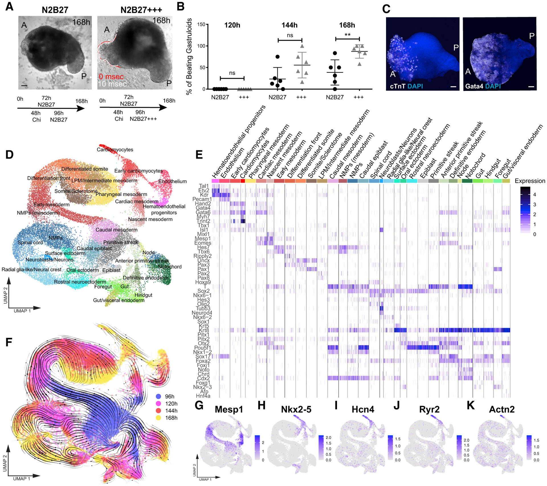
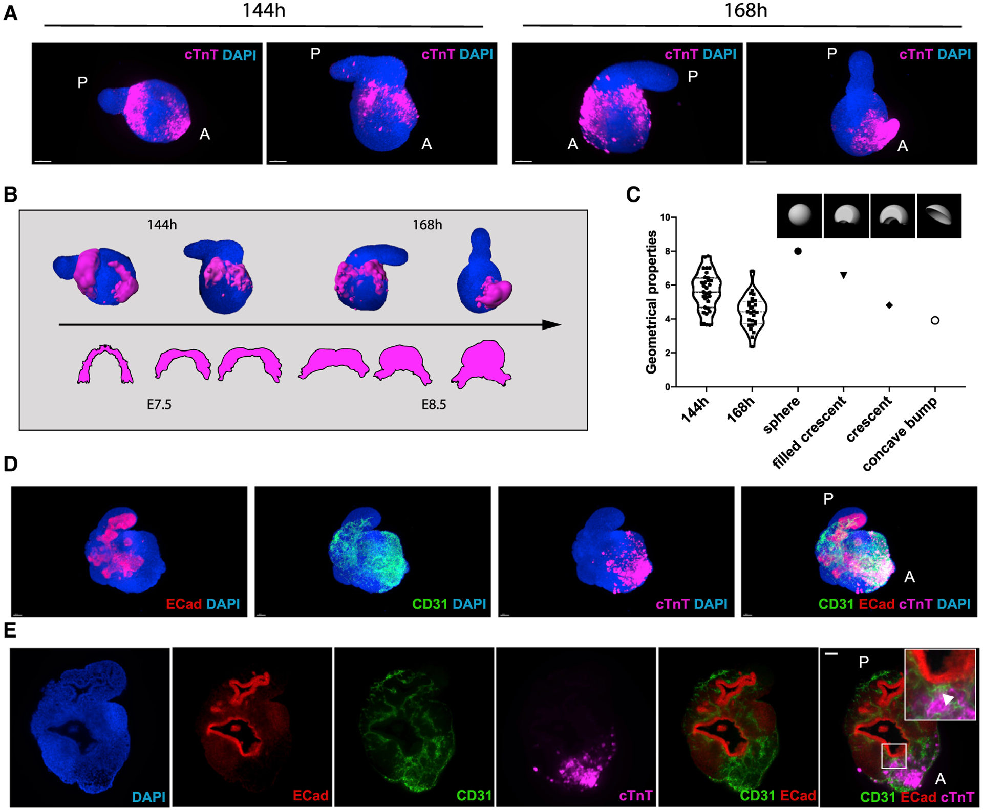
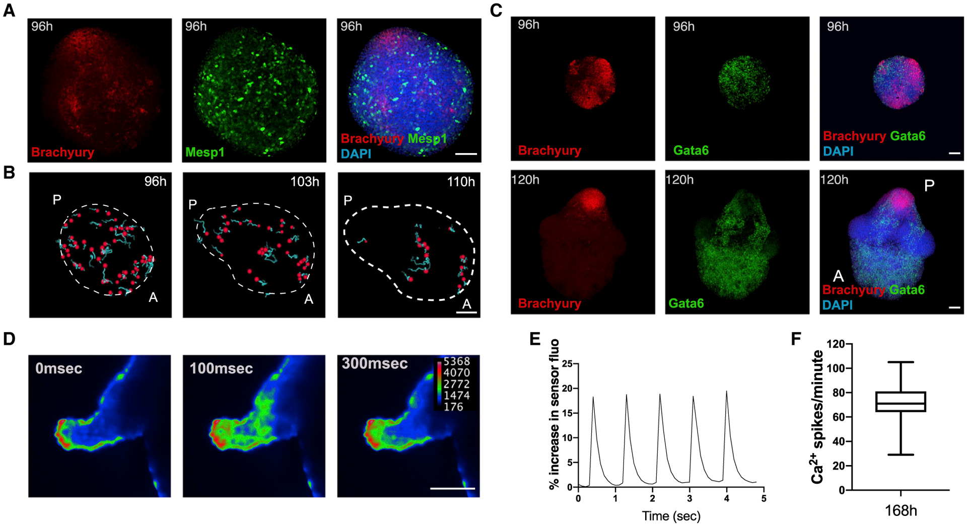
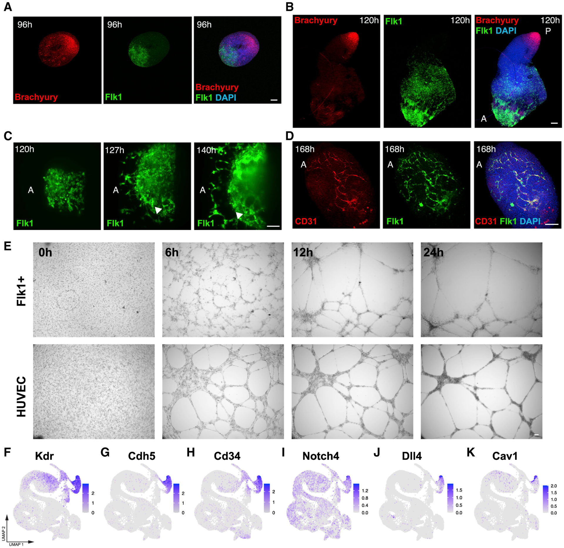
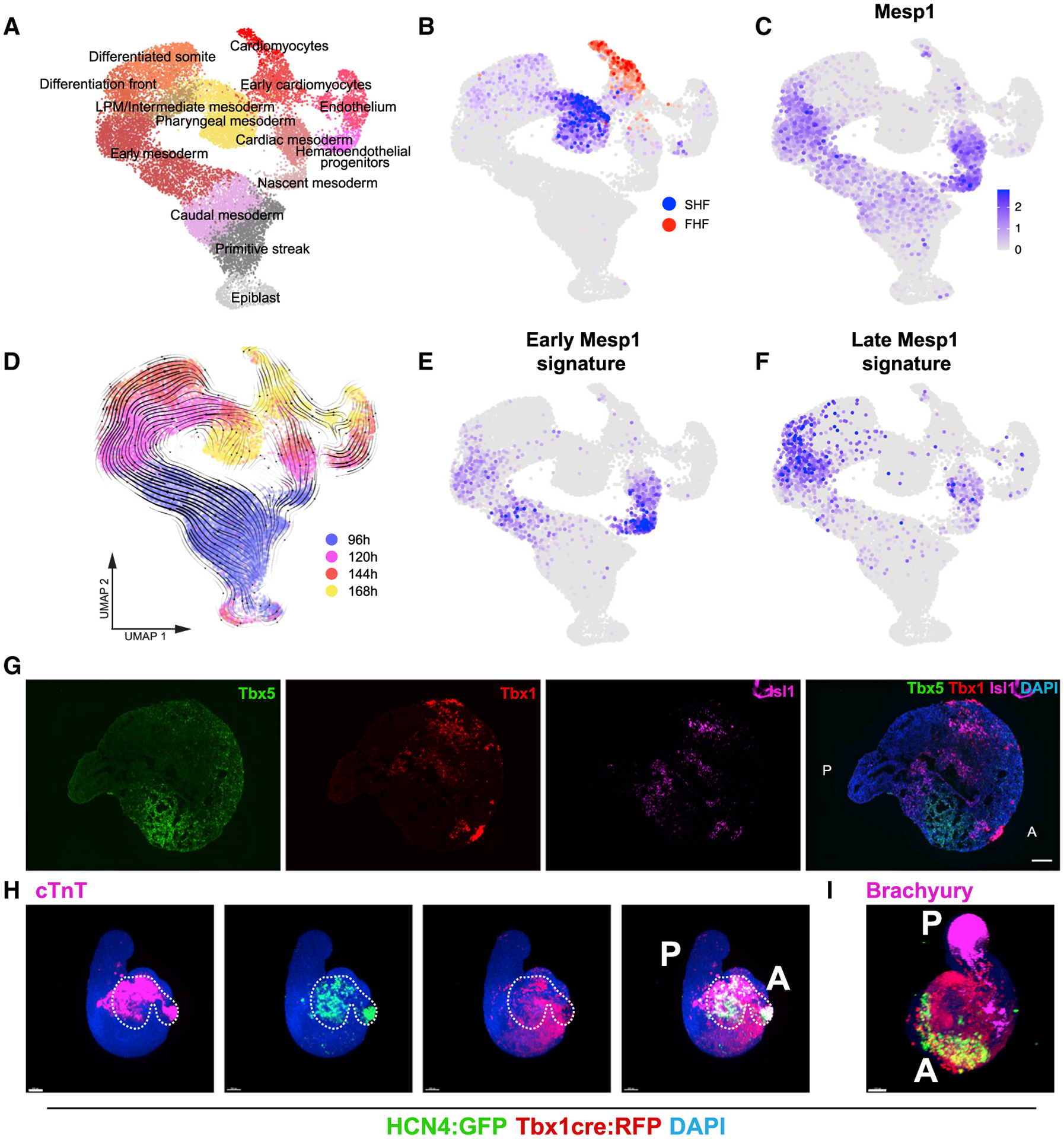
Organoids are powerful models for studying tissue development, physiology, and disease. However, current culture systems disrupt the inductive tissue-tissue interactions needed for the complex morphogenetic processes of native organogenesis. Here, we show that mouse embryonic stem cells (mESCs) can be coaxed to robustly undergo fundamental steps of early heart organogenesis with an in-vivo-like spatiotemporal fidelity. These axially patterned embryonic organoids (gastruloids) mimic embryonic development and support the generation of cardiovascular progenitors, including first and second heart fields. The cardiac progenitors self-organize into an anterior domain reminiscent of a cardiac crescent before forming a beating cardiac tissue near a putative primitive gut-like tube, from which it is separated by an endocardial-like layer. These findings unveil the surprising morphogenetic potential of mESCs to execute key aspects of organogenesis through the coordinated development of multiple tissues. This platform could be an excellent tool for studying heart development in unprecedented detail and throughput.
类器官是研究组织发育、生理学和疾病的强大模型。然而,目前的培养系统破坏了诱导组织-组织相互作用的需要,而这种相互作用是原生器官发生的复杂形态发生过程所必需的。在这里,我们展示了小鼠胚胎干细胞(mESCs)可以被诱导以具有类似于体内的时空保真度来进行早期心脏器官发生的基本步骤。这些轴向模式化的胚胎类器官(原肠胚)模拟胚胎发育,并支持心血管祖细胞的产生,包括第一和第二心脏场。心脏祖细胞在形成类似于原始肠样管的心脏新月形之前,会自我组织成一个前域,然后在一个假定的原始肠样管附近形成跳动的心脏组织,其间由心内膜样层隔开。这些发现揭示了 mESCs 通过多个组织的协调发育来执行器官发生关键方面的惊人形态发生潜力。该平台可能是一个极好的工具,可以以前所未有的细节和通量研究心脏发育。