Zhao Ming-Tao, Ye Shiqiao, Su Juan, Garg Vidu
Center for Cardiovascular Research, The Abigail Wexner Research Institute, Nationwide Children's Hospital, Columbus, OH, United States.
The Heart Center, Nationwide Children's Hospital, Columbus, OH, United States.
Front Cell Dev Biol. 2020 Oct 15;8:594226. doi: 10.3389/fcell.2020.594226. eCollection 2020.
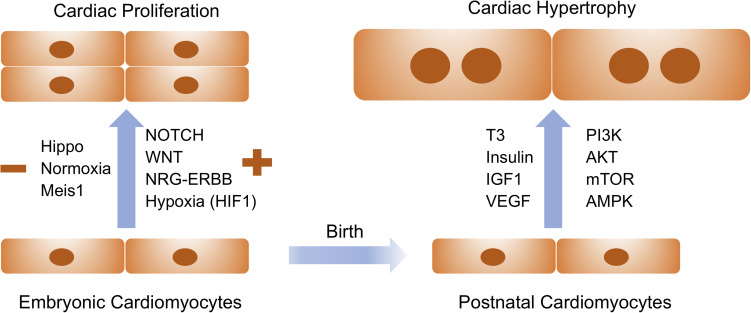
In the past few decades, cardiac regeneration has been the central target for restoring the injured heart. In mammals, cardiomyocytes are terminally differentiated and rarely divide during adulthood. Embryonic and fetal cardiomyocytes undergo robust proliferation to form mature heart chambers in order to accommodate the increased workload of a systemic circulation. In contrast, postnatal cardiomyocytes stop dividing and initiate hypertrophic growth by increasing the size of the cardiomyocyte when exposed to increased workload. Extracellular and intracellular signaling pathways control embryonic cardiomyocyte proliferation and postnatal cardiac hypertrophy. Harnessing these pathways could be the future focus for stimulating endogenous cardiac regeneration in response to various pathological stressors. Meanwhile, patient-specific cardiomyocytes derived from autologous induced pluripotent stem cells (iPSCs) could become the major exogenous sources for replenishing the damaged myocardium. Human iPSC-derived cardiomyocytes (iPSC-CMs) are relatively immature and have the potential to increase the population of cells that advance to physiological hypertrophy in the presence of extracellular stimuli. In this review, we discuss how cardiac proliferation and maturation are regulated during embryonic development and postnatal growth, and explore how patient iPSC-CMs could serve as the future seed cells for cardiac cell replacement therapy.
在过去几十年里,心脏再生一直是修复受损心脏的核心目标。在哺乳动物中,心肌细胞在成年期终末分化且很少分裂。胚胎期和胎儿期的心肌细胞会经历旺盛的增殖以形成成熟的心脏腔室,从而适应体循环增加的工作量。相比之下,出生后的心肌细胞停止分裂,并在面临工作量增加时通过增大细胞体积启动肥厚性生长。细胞外和细胞内信号通路控制着胚胎期心肌细胞的增殖以及出生后心脏的肥厚。利用这些信号通路可能是未来针对各种病理应激源刺激内源性心脏再生的重点。同时,源自自体诱导多能干细胞(iPSC)的患者特异性心肌细胞可能会成为补充受损心肌的主要外源性来源。人iPSC衍生的心肌细胞(iPSC-CM)相对不成熟,并且在存在细胞外刺激的情况下有增加向生理性肥厚发展的细胞数量的潜力。在这篇综述中,我们讨论了胚胎发育和出生后生长过程中心脏增殖和成熟是如何被调控的,并探讨了患者iPSC-CM如何能够作为未来心脏细胞替代疗法的种子细胞。