Rezk Abdelrahman I, Kim Kyung-Suk, Kim Cheol Sang
Department of Bionanosystem Engineering, Graduate School, Jeonbuk National University, Jeonju, Jeonbuk 561-756, Korea.
Department of Bionanotechnology and Bioconvergence Engineering, Graduate School, Jeonbuk National University, Jeonju, Jeonbuk 561-756, Korea.
Polymers (Basel). 2020 Nov 12;12(11):2667. doi: 10.3390/polym12112667.
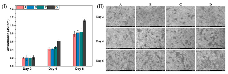
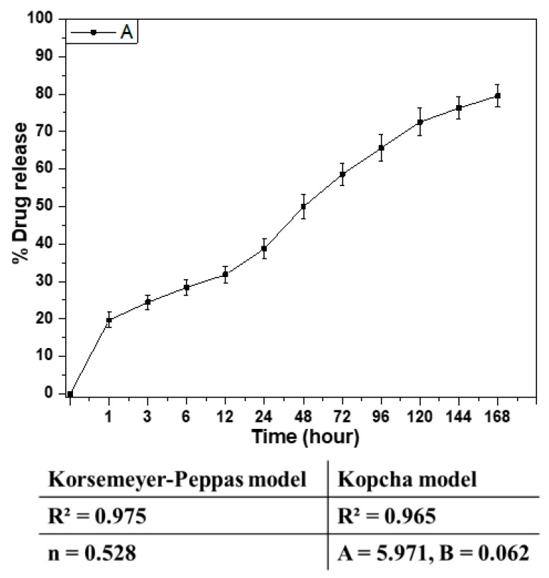
Herein, we report a drug eluting scaffold composed of a composite nanofibers of poly(ε-caprolactone) (PCL) and poly(glycerol sebacate) (PGS) loaded with Hydroxyapatite nanoparticles (HANPs) and simvastatin (SIM) mimicking the bone extracellular matrix (ECM) to improve bone cell proliferation and regeneration process. Indeed, the addition of PGS results in a slight increase in the average fiber diameter compared to PCL. However, the presence of HANPs in the composite nanofibers induced a greater fiber diameter distribution, without significantly changing the average fiber diameter. The in vitro drug release result revealed that the sustained release of SIM from the composite nanofiber obeying the Korsemeyer-Peppas and Kpocha models revealing a non-Fickian diffusion mechanism and the release mechanism follows diffusion rather than polymer erosion. Biomineralization assessment of the nanofibers was carried out in simulated body fluid (SBF). SEM and EDS analysis confirmed nucleation of the hydroxyapatite layer on the surface of the composite nanofibers mimicking the natural apatite layer. Moreover, in vitro studies revealed that the PCL-PGS-HA displayed better cell proliferation and adhesion compared to the control sample, hence improving the regeneration process. This suggests that the fabricated PCL-PGS-HA could be a promising future scaffold for control drug delivery and bone tissue regeneration application.
在此,我们报道了一种药物洗脱支架,其由聚(ε-己内酯)(PCL)和聚(癸二酸甘油酯)(PGS)的复合纳米纤维组成,负载有羟基磷灰石纳米颗粒(HANPs)和辛伐他汀(SIM),模仿骨细胞外基质(ECM)以改善骨细胞增殖和再生过程。实际上,与PCL相比,PGS的添加导致平均纤维直径略有增加。然而,复合纳米纤维中HANPs的存在导致更大的纤维直径分布,而平均纤维直径没有显著变化。体外药物释放结果表明,SIM从复合纳米纤维中的持续释放符合Korsemeyer-Peppas和Kpocha模型,揭示了非菲克扩散机制,且释放机制遵循扩散而非聚合物侵蚀。在模拟体液(SBF)中对纳米纤维进行了生物矿化评估。扫描电子显微镜(SEM)和能谱分析(EDS)证实了在复合纳米纤维表面有羟基磷灰石层的成核,模仿了天然磷灰石层。此外,体外研究表明,与对照样品相比,PCL-PGS-HA表现出更好的细胞增殖和粘附,从而改善了再生过程。这表明所制备的PCL-PGS-HA可能是未来用于控制药物递送和骨组织再生应用的有前途的支架。