Ball Gareth, Seidlitz Jakob, O'Muircheartaigh Jonathan, Dimitrova Ralica, Fenchel Daphna, Makropoulos Antonios, Christiaens Daan, Schuh Andreas, Passerat-Palmbach Jonathan, Hutter Jana, Cordero-Grande Lucilio, Hughes Emer, Price Anthony, Hajnal Jo V, Rueckert Daniel, Robinson Emma C, Edwards A David
Developmental Imaging, Murdoch Children's Research Institute, Melbourne, Australia.
Centre for the Developing Brain, Department of Perinatal Imaging & Health, King's College London, United Kingdom.
PLoS Biol. 2020 Nov 23;18(11):e3000976. doi: 10.1371/journal.pbio.3000976. eCollection 2020 Nov.
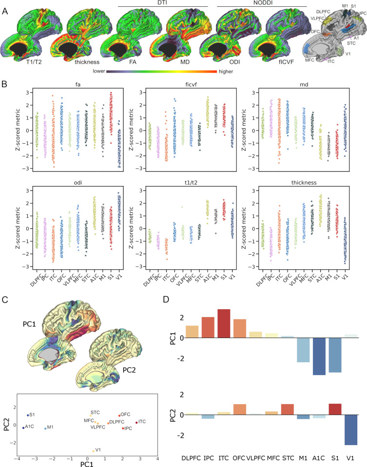
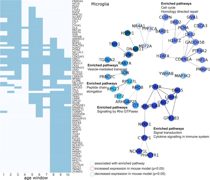
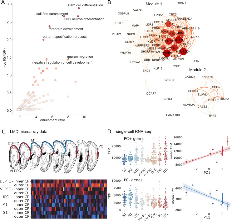
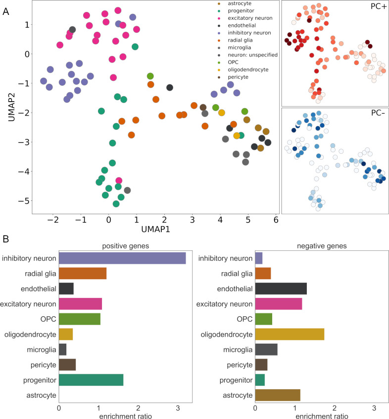
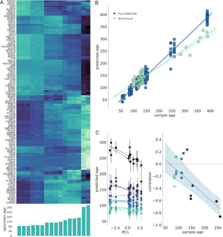
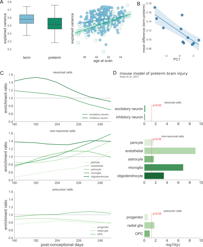
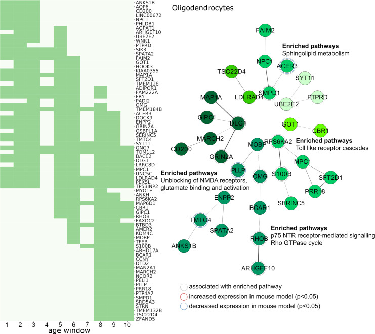
Interruption to gestation through preterm birth can significantly impact cortical development and have long-lasting adverse effects on neurodevelopmental outcome. We compared cortical morphology captured by high-resolution, multimodal magnetic resonance imaging (MRI) in n = 292 healthy newborn infants (mean age at birth = 39.9 weeks) with regional patterns of gene expression in the fetal cortex across gestation (n = 156 samples from 16 brains, aged 12 to 37 postconceptional weeks [pcw]). We tested the hypothesis that noninvasive measures of cortical structure at birth mirror areal differences in cortical gene expression across gestation, and in a cohort of n = 64 preterm infants (mean age at birth = 32.0 weeks), we tested whether cortical alterations observed after preterm birth were associated with altered gene expression in specific developmental cell populations. Neonatal cortical structure was aligned to differential patterns of cell-specific gene expression in the fetal cortex. Principal component analysis (PCA) of 6 measures of cortical morphology and microstructure showed that cortical regions were ordered along a principal axis, with primary cortex clearly separated from heteromodal cortex. This axis was correlated with estimated tissue maturity, indexed by differential expression of genes expressed by progenitor cells and neurons, and engaged in stem cell differentiation, neuron migration, and forebrain development. Preterm birth was associated with altered regional MRI metrics and patterns of differential gene expression in glial cell populations. The spatial patterning of gene expression in the developing cortex was thus mirrored by regional variation in cortical morphology and microstructure at term, and this was disrupted by preterm birth. This work provides a framework to link molecular mechanisms to noninvasive measures of cortical development in early life and highlights novel pathways to injury in neonatal populations at increased risk of neurodevelopmental disorder.
早产导致的妊娠中断会显著影响皮质发育,并对神经发育结局产生长期的不良影响。我们比较了292名健康新生儿(出生时平均年龄 = 39.9周)通过高分辨率多模态磁共振成像(MRI)获取的皮质形态,以及整个孕期胎儿皮质中的基因表达区域模式(来自16个大脑的156个样本,孕龄为12至37周[pcw])。我们检验了这样一个假设,即出生时皮质结构的非侵入性测量反映了整个孕期皮质基因表达的区域差异,并且在一个由64名早产儿组成的队列(出生时平均年龄 = 32.0周)中,我们检验了早产之后观察到的皮质改变是否与特定发育细胞群体中基因表达的改变有关。新生儿皮质结构与胎儿皮质中细胞特异性基因表达的差异模式相匹配。对6种皮质形态和微结构测量指标进行主成分分析(PCA)表明,皮质区域沿着一个主轴排列,初级皮质与异模态皮质明显分开。这个轴与估计的组织成熟度相关,通过祖细胞和神经元表达的基因的差异表达来衡量,并且参与干细胞分化、神经元迁移和前脑发育。早产与神经胶质细胞群体中区域MRI指标的改变以及差异基因表达模式有关。因此,发育中皮质基因表达的空间模式在足月时由皮质形态和微结构的区域差异所反映,而这被早产所破坏。这项工作提供了一个框架,将分子机制与生命早期皮质发育的非侵入性测量联系起来,并突出了在神经发育障碍风险增加的新生儿群体中损伤的新途径。