• 文献检索
  • 文档翻译
  • 深度研究
  • 学术资讯
  • Suppr Zotero 插件Zotero 插件
  • 邀请有礼
  • 套餐&价格
  • 历史记录
应用&插件
Suppr Zotero 插件Zotero 插件浏览器插件Mac 客户端Windows 客户端微信小程序
定价
高级版会员购买积分包购买API积分包
服务
文献检索文档翻译深度研究API 文档MCP 服务
关于我们
关于 Suppr公司介绍联系我们用户协议隐私条款
关注我们

Suppr 超能文献

核心技术专利:CN118964589B侵权必究
粤ICP备2023148730 号-1Suppr @ 2026

文献检索

告别复杂PubMed语法,用中文像聊天一样搜索,搜遍4000万医学文献。AI智能推荐,让科研检索更轻松。

立即免费搜索

文件翻译

保留排版,准确专业,支持PDF/Word/PPT等文件格式,支持 12+语言互译。

免费翻译文档

深度研究

AI帮你快速写综述,25分钟生成高质量综述,智能提取关键信息,辅助科研写作。

立即免费体验

手工特征与卷积神经网络在肝脏磁共振图像充分性评估中的比较。

Comparison of handcrafted features and convolutional neural networks for liver MR image adequacy assessment.

机构信息

Division of Biostatistics, Department of Family Medicine and Public Health, University of California San Diego, La Jolla, 92093, USA.

Department of Mathematics and Statistics, San Diego State University, San Diego, CA, 92182, USA.

出版信息

Sci Rep. 2020 Nov 23;10(1):20336. doi: 10.1038/s41598-020-77264-y.

DOI:10.1038/s41598-020-77264-y
PMID:33230152
原文链接:https://pmc.ncbi.nlm.nih.gov/articles/PMC7683555/
Abstract

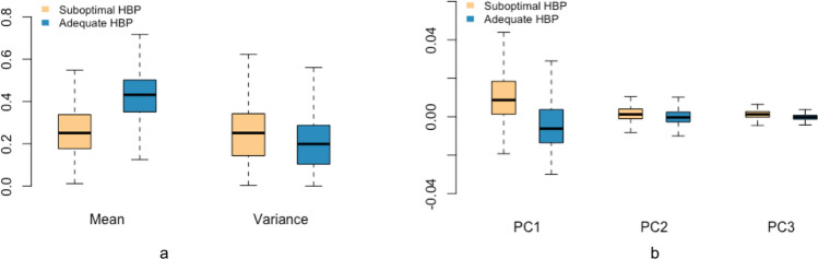
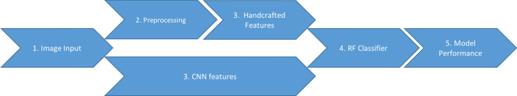
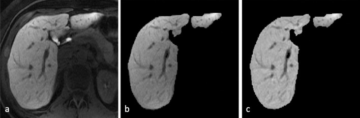
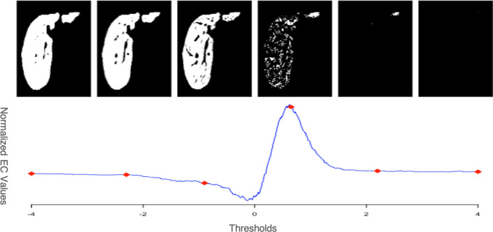
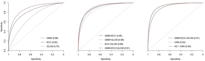
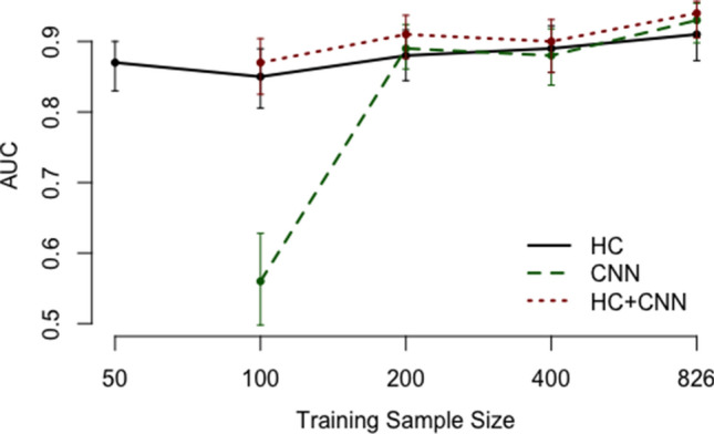
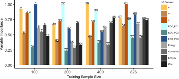
We propose a random forest classifier for identifying adequacy of liver MR images using handcrafted (HC) features and deep convolutional neural networks (CNNs), and analyze the relative role of these two components in relation to the training sample size. The HC features, specifically developed for this application, include Gaussian mixture models, Euler characteristic curves and texture analysis. Using HC features outperforms the CNN for smaller sample sizes and with increased interpretability. On the other hand, with enough training data, the combined classifier outperforms the models trained with HC features or CNN features alone. These results illustrate the added value of HC features with respect to CNNs, especially when insufficient data is available, as is often found in clinical studies.

摘要

我们提出了一种随机森林分类器,用于使用手工制作(HC)特征和深度卷积神经网络(CNNs)来识别肝脏磁共振图像的充分性,并分析这两个组件相对于训练样本大小的相对作用。HC 特征是专门为此应用开发的,包括高斯混合模型、欧拉特征曲线和纹理分析。在较小的样本量下,使用 HC 特征比 CNN 表现更好,并且具有更高的可解释性。另一方面,随着训练数据的增加,组合分类器的性能优于仅使用 HC 特征或 CNN 特征训练的模型。这些结果说明了 HC 特征相对于 CNN 的附加价值,特别是在数据不足的情况下,这种情况在临床研究中经常出现。

https://cdn.ncbi.nlm.nih.gov/pmc/blobs/68a1/7683555/044c1d635f56/41598_2020_77264_Fig10_HTML.jpg
https://cdn.ncbi.nlm.nih.gov/pmc/blobs/68a1/7683555/d0bf109ec035/41598_2020_77264_Fig1_HTML.jpg
https://cdn.ncbi.nlm.nih.gov/pmc/blobs/68a1/7683555/541eeac8dc72/41598_2020_77264_Fig2_HTML.jpg
https://cdn.ncbi.nlm.nih.gov/pmc/blobs/68a1/7683555/58a7a30440a0/41598_2020_77264_Fig3_HTML.jpg
https://cdn.ncbi.nlm.nih.gov/pmc/blobs/68a1/7683555/f74fc9127635/41598_2020_77264_Fig4_HTML.jpg
https://cdn.ncbi.nlm.nih.gov/pmc/blobs/68a1/7683555/e0f06d4a18d2/41598_2020_77264_Fig5_HTML.jpg
https://cdn.ncbi.nlm.nih.gov/pmc/blobs/68a1/7683555/3804bce6e063/41598_2020_77264_Fig6_HTML.jpg
https://cdn.ncbi.nlm.nih.gov/pmc/blobs/68a1/7683555/84ed9f379579/41598_2020_77264_Fig7_HTML.jpg
https://cdn.ncbi.nlm.nih.gov/pmc/blobs/68a1/7683555/9d1b85e6d7a4/41598_2020_77264_Fig8_HTML.jpg
https://cdn.ncbi.nlm.nih.gov/pmc/blobs/68a1/7683555/213686a5dafd/41598_2020_77264_Fig9_HTML.jpg
https://cdn.ncbi.nlm.nih.gov/pmc/blobs/68a1/7683555/044c1d635f56/41598_2020_77264_Fig10_HTML.jpg

相似文献

1
Comparison of handcrafted features and convolutional neural networks for liver MR image adequacy assessment.手工特征与卷积神经网络在肝脏磁共振图像充分性评估中的比较。
Sci Rep. 2020 Nov 23;10(1):20336. doi: 10.1038/s41598-020-77264-y.
2
Deep learning approaches using 2D and 3D convolutional neural networks for generating male pelvic synthetic computed tomography from magnetic resonance imaging.使用 2D 和 3D 卷积神经网络从磁共振成像生成男性骨盆合成 CT 的深度学习方法。
Med Phys. 2019 Sep;46(9):3788-3798. doi: 10.1002/mp.13672. Epub 2019 Jul 26.
3
Mitosis detection in breast cancer pathology images by combining handcrafted and convolutional neural network features.通过结合手工制作的特征和卷积神经网络特征来检测乳腺癌病理图像中的有丝分裂。
J Med Imaging (Bellingham). 2014 Oct;1(3):034003. doi: 10.1117/1.JMI.1.3.034003. Epub 2014 Oct 10.
4
Co-trained convolutional neural networks for automated detection of prostate cancer in multi-parametric MRI.基于多参数 MRI 的协同训练卷积神经网络在前列腺癌自动检测中的应用
Med Image Anal. 2017 Dec;42:212-227. doi: 10.1016/j.media.2017.08.006. Epub 2017 Aug 24.
5
Automated diagnosis of prostate cancer in multi-parametric MRI based on multimodal convolutional neural networks.基于多模态卷积神经网络的多参数磁共振成像中前列腺癌的自动诊断
Phys Med Biol. 2017 Jul 24;62(16):6497-6514. doi: 10.1088/1361-6560/aa7731.
6
Brain tumor classification for MR images using transfer learning and fine-tuning.基于迁移学习和微调的磁共振图像脑肿瘤分类。
Comput Med Imaging Graph. 2019 Jul;75:34-46. doi: 10.1016/j.compmedimag.2019.05.001. Epub 2019 May 18.
7
Deep learning for patient-specific quality assurance: Identifying errors in radiotherapy delivery by radiomic analysis of gamma images with convolutional neural networks.深度学习在个体化质量保证中的应用:通过卷积神经网络对伽马图像的放射组学分析识别放射治疗中的误差。
Med Phys. 2019 Feb;46(2):456-464. doi: 10.1002/mp.13338. Epub 2018 Dec 28.
8
Study of the Application of Deep Convolutional Neural Networks (CNNs) in Processing Sensor Data and Biomedical Images.深度学习卷积神经网络(CNNs)在传感器数据和生物医学图像处理中的应用研究。
Sensors (Basel). 2019 Aug 17;19(16):3584. doi: 10.3390/s19163584.
9
Patient-specific and global convolutional neural networks for robust automatic liver tumor delineation in follow-up CT studies.用于在随访 CT 研究中进行稳健自动肝肿瘤勾画的基于患者特定和全局卷积神经网络。
Med Biol Eng Comput. 2018 Sep;56(9):1699-1713. doi: 10.1007/s11517-018-1803-6. Epub 2018 Mar 10.
10
Neonatal Seizure Detection Using Deep Convolutional Neural Networks.使用深度卷积神经网络进行新生儿癫痫发作检测。
Int J Neural Syst. 2019 May;29(4):1850011. doi: 10.1142/S0129065718500119. Epub 2018 Apr 2.

引用本文的文献

1
When liver disease diagnosis encounters deep learning: Analysis, challenges, and prospects.当肝脏疾病诊断遇上深度学习:分析、挑战与展望。
ILIVER. 2023 Mar 4;2(1):73-87. doi: 10.1016/j.iliver.2023.02.002. eCollection 2023 Mar.
2
Soft Bioelectronic Interfaces for Continuous Peripheral Neural Signal Recording and Robust Cross-Subject Decoding.用于连续外周神经信号记录和稳健跨受试者解码的柔性生物电子接口。
Adv Sci (Weinh). 2025 Sep;12(33):e14732. doi: 10.1002/advs.202414732. Epub 2025 May 28.
3
The role of AI for MRI-analysis in multiple sclerosis-A brief overview.

本文引用的文献

1
Automated CT and MRI Liver Segmentation and Biometry Using a Generalized Convolutional Neural Network.使用广义卷积神经网络的肝脏自动CT和MRI分割与生物测量
Radiol Artif Intell. 2019 Mar;1(2). doi: 10.1148/ryai.2019180022. Epub 2019 Mar 27.
2
Integration of Random Forest Classifiers and Deep Convolutional Neural Networks for Classification and Biomolecular Modeling of Cancer Driver Mutations.随机森林分类器与深度卷积神经网络的集成用于癌症驱动突变的分类和生物分子建模
Front Mol Biosci. 2019 Jun 11;6:44. doi: 10.3389/fmolb.2019.00044. eCollection 2019.
3
Automated estimation of image quality for coronary computed tomographic angiography using machine learning.
人工智能在多发性硬化症磁共振成像分析中的作用——简要概述。
Front Artif Intell. 2025 Apr 8;8:1478068. doi: 10.3389/frai.2025.1478068. eCollection 2025.
4
A fully automated, expert-perceptive image quality assessment system for whole-body [18F]FDG PET/CT.一种用于全身[18F]FDG PET/CT的全自动、专家感知图像质量评估系统。
EJNMMI Res. 2025 Apr 18;15(1):42. doi: 10.1186/s13550-025-01238-2.
5
Deep phenotyping platform for microscopic plant-pathogen interactions.用于微观植物-病原体相互作用的深度表型分析平台。
Front Plant Sci. 2025 Feb 3;16:1462694. doi: 10.3389/fpls.2025.1462694. eCollection 2025.
6
Extreme learning machine for identifying soil-dwelling microorganisms cultivated on agar media.用于识别在琼脂培养基上培养的土壤微生物的极限学习机。
Sci Rep. 2024 Dec 28;14(1):31034. doi: 10.1038/s41598-024-82174-4.
7
STEFF: Spatio-temporal EfficientNet for dynamic texture classification in outdoor scenes.STEFF:用于室外场景动态纹理分类的时空高效神经网络
Heliyon. 2024 Feb 5;10(3):e25360. doi: 10.1016/j.heliyon.2024.e25360. eCollection 2024 Feb 15.
8
Media Forensics Considerations on DeepFake Detection with Hand-Crafted Features.基于手工特征的深度伪造检测中的媒体取证考量
J Imaging. 2021 Jul 1;7(7):108. doi: 10.3390/jimaging7070108.
9
Deep learning in bioinformatics.生物信息学中的深度学习。
Turk J Biol. 2023 Dec 18;47(6):366-382. doi: 10.55730/1300-0152.2671. eCollection 2023.
10
Automatic and real-time tissue sensing for autonomous intestinal anastomosis using hybrid MLP-DC-CNN classifier-based optical coherence tomography.使用基于混合MLP-DC-CNN分类器的光学相干断层扫描技术实现自主肠道吻合的自动实时组织传感。
Biomed Opt Express. 2024 Mar 25;15(4):2543-2560. doi: 10.1364/BOE.521652. eCollection 2024 Apr 1.
使用机器学习自动评估冠状动脉 CT 血管造影的图像质量。
Eur Radiol. 2018 Sep;28(9):4018-4026. doi: 10.1007/s00330-018-5348-8. Epub 2018 Mar 23.
4
Machine Learning in Radiology: Applications Beyond Image Interpretation.机器学习在放射学中的应用:超越图像解读的应用。
J Am Coll Radiol. 2018 Feb;15(2):350-359. doi: 10.1016/j.jacr.2017.09.044. Epub 2017 Nov 17.
5
Computational Radiomics System to Decode the Radiographic Phenotype.用于解码影像学表型的计算放射组学系统
Cancer Res. 2017 Nov 1;77(21):e104-e107. doi: 10.1158/0008-5472.CAN-17-0339.
6
Automated image quality evaluation of T -weighted liver MRI utilizing deep learning architecture.利用深度学习架构进行 T 加权肝脏 MRI 的自动图像质量评估。
J Magn Reson Imaging. 2018 Mar;47(3):723-728. doi: 10.1002/jmri.25779. Epub 2017 Jun 3.
7
Automated Quality Assessment of Structural Magnetic Resonance Brain Images Based on a Supervised Machine Learning Algorithm.基于监督式机器学习算法的脑部结构磁共振图像自动质量评估
Front Neuroinform. 2016 Dec 19;10:52. doi: 10.3389/fninf.2016.00052. eCollection 2016.
8
Machine learning approaches in medical image analysis: From detection to diagnosis.机器学习在医学图像分析中的应用:从检测到诊断。
Med Image Anal. 2016 Oct;33:94-97. doi: 10.1016/j.media.2016.06.032. Epub 2016 Jun 23.
9
Focal liver lesions: Practical magnetic resonance imaging approach.肝脏局灶性病变:实用磁共振成像方法
World J Hepatol. 2015 Aug 8;7(16):1987-2008. doi: 10.4254/wjh.v7.i16.1987.
10
Hepatitis C related chronic liver cirrhosis: feasibility of texture analysis of MR images for classification of fibrosis stage and necroinflammatory activity grade.丙型肝炎相关慢性肝硬化:磁共振图像纹理分析用于纤维化分期和坏死性炎症活动度分级的可行性
PLoS One. 2015 Mar 5;10(3):e0118297. doi: 10.1371/journal.pone.0118297. eCollection 2015.