Division of Molecular and Developmental Biology, Institute of Medical Science, University of Tokyo, Tokyo, Japan.
Department of Molecular Oncology, Graduate School of Medicine, Kyoto University, Kyoto, Japan.
Invest Ophthalmol Vis Sci. 2020 Nov 2;61(13):34. doi: 10.1167/iovs.61.13.34.
The role of activated retinal microglia in driving retinal degeneration has been implicated in a number of in vivo disease models. Here, we investigated the primary consequences of microglial activation by the specific expression of constitutively active Ras in microglia in a transgenic mouse model before the onset of any degenerative changes in the retina.
The double transgenic lines CAG-LSL-RasV12-IRES-EGFP; Cx3cr1CreER/+ (Cx3cr1-RasV12 mice) and CAG-LSL-EGFP; Cx3cr1CreER_+ (control mice) were generated. The expression of RasV12 was induced in microglia by tamoxifen administration, and the retinas were examined by immunohistochemistry of frozen sections, RT-qPCR, and live imaging.
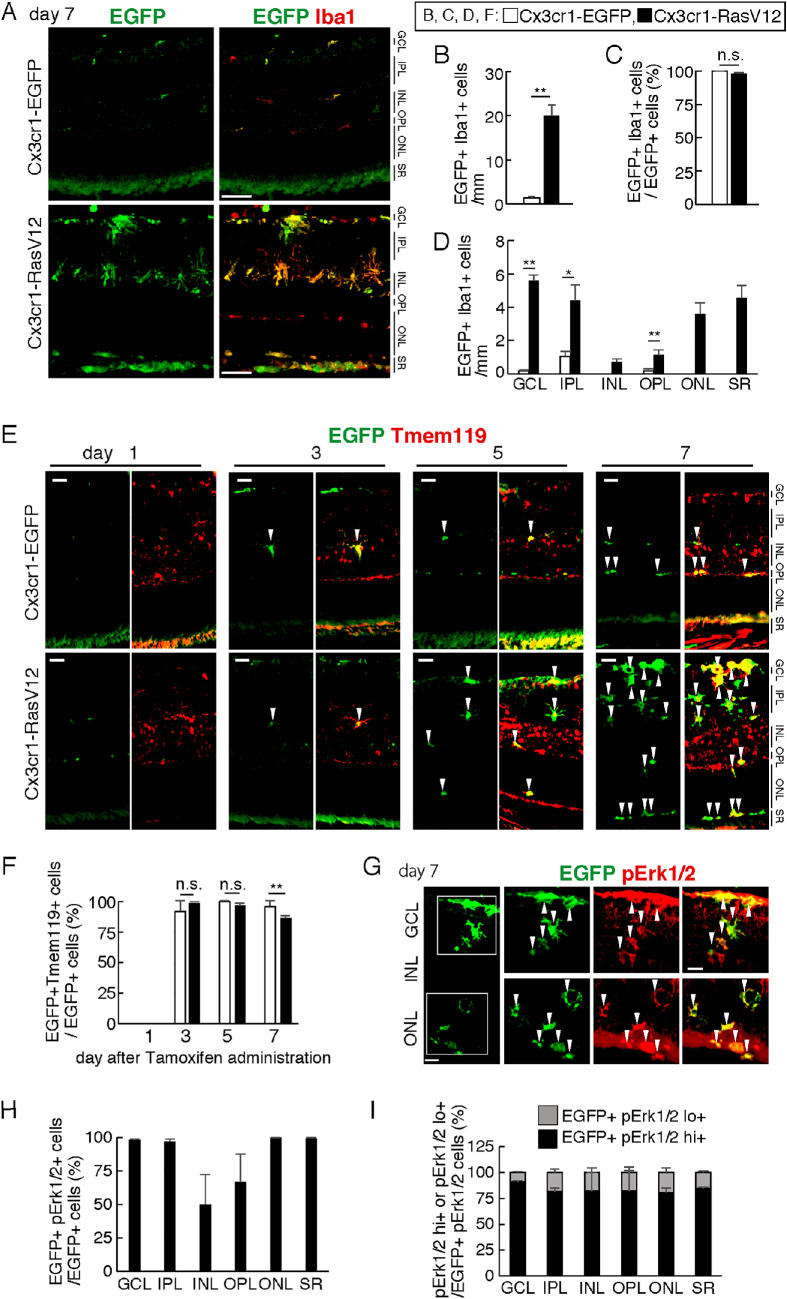
RasV12 expression in retinal microglial cells promoted cell proliferation, cytokine expression, and phagocytosis. RasV12-expressing microglia migrated toward the inner and outer layers of the retina. Examination of glial fibrillary acidic protein (GFAP) expression revealed activation of Müller glia in the retina. We also observed loss of the photoreceptors in the outer nuclear layer in close proximity to microglial cells. However, no significant neurodegeneration was detected in the inner nuclear layer (INL) or ganglion cell layer (GCL). The morphology of RasV12-expressing microglia in the GCL and INL retained more ramified features compared with the predominantly-ameboid morphology found in outer retinal microglia.
The expression of RasV12 is sufficient to activate microglia and lead to photoreceptor degeneration. Neurons in the inner side of the retina were not damaged by the RasV12-activated microglia, suggesting that microenvironment cues may modulate the microglial phenotypic features and effects of microglial activation.
在许多体内疾病模型中,激活的视网膜小胶质细胞在驱动视网膜变性中的作用已被牵连。在这里,我们在视网膜发生任何退行性变化之前,通过在转基因小鼠模型中特异性表达组成型激活的 Ras 来研究小胶质细胞激活的主要后果。
生成了双转基因系 CAG-LSL-RasV12-IRES-EGFP;Cx3cr1CreER/+(Cx3cr1-RasV12 小鼠)和 CAG-LSL-EGFP;Cx3cr1CreER_+(对照小鼠)。通过给予他莫昔芬诱导 RasV12 在小胶质细胞中的表达,并通过对冷冻切片、RT-qPCR 和活体成像进行免疫组织化学染色来检查视网膜。
视网膜小胶质细胞中 RasV12 的表达促进了细胞增殖、细胞因子表达和吞噬作用。RasV12 表达的小胶质细胞向视网膜的内、外两层迁移。胶质纤维酸性蛋白 (GFAP) 表达的检查显示视网膜中的 Müller 胶质细胞被激活。我们还观察到靠近小胶质细胞的外核层中的光感受器丧失。然而,在内核层 (INL) 或神经节细胞层 (GCL) 中未检测到明显的神经退行性变。与在外视网膜小胶质细胞中发现的主要阿米巴样形态相比,RasV12 表达的小胶质细胞在 GCL 和 INL 中的形态保持更多的分支特征。
RasV12 的表达足以激活小胶质细胞并导致光感受器变性。内视网膜侧的神经元未被 RasV12 激活的小胶质细胞损伤,这表明微环境线索可能调节小胶质细胞表型特征和小胶质细胞激活的影响。