Jiangsu Key Laboratory of Molecular and Functional Imaging, Department of Radiology, Zhongda Hospital, Medical School of Southeast University, Nanjing 210009, People's Republic of China.
Int J Nanomedicine. 2020 Nov 17;15:9011-9023. doi: 10.2147/IJN.S271519. eCollection 2020.
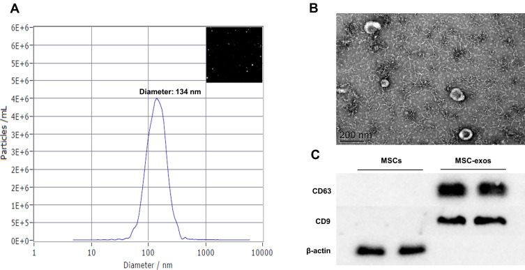
Mesenchymal stem cell-derived exosomes (MSC-exos) are considered an important restorative treatment for ischemic stroke. However, the migration ability and survival of exogenous MSC-exos remain unclear. Here, we investigated whether MSC-exos migrate into the ischemic brain and play a protective role against ischemic stroke.
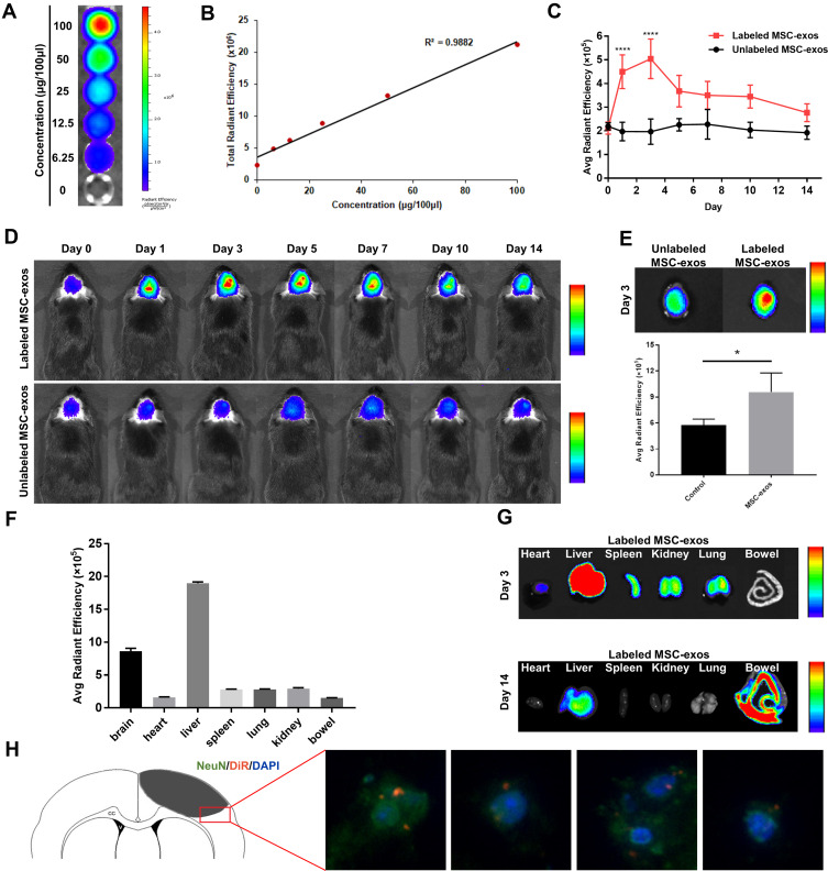
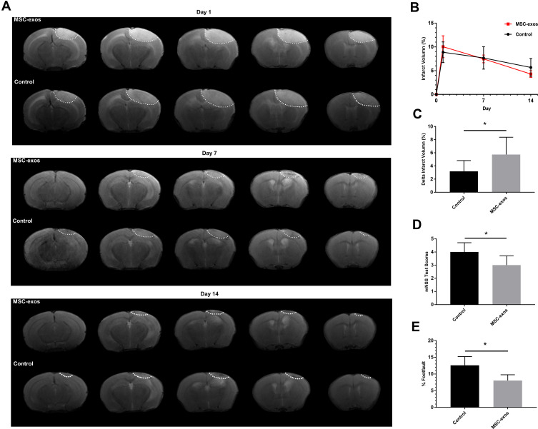
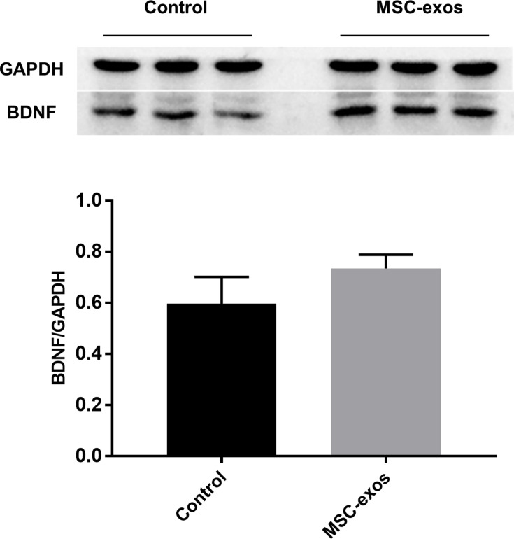
MSC-exos labeled with DiR were injected intravenously into mice with ischemic stroke. Near-infrared fluorescence (NIRF) images were obtained on days 0, 1, 3, 5, 7, 10, and 14, and magnetic resonance (MR) images were obtained on days 1, 7 and 14. On day 14, the functional outcomes, angiogenesis, neurogenesis, and white matter remodeling were assessed, and Western blot assays were performed.
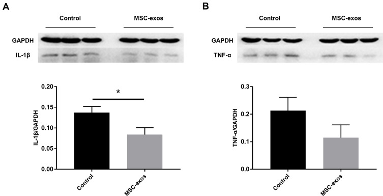
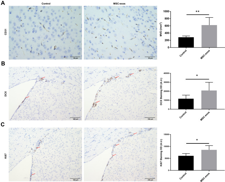
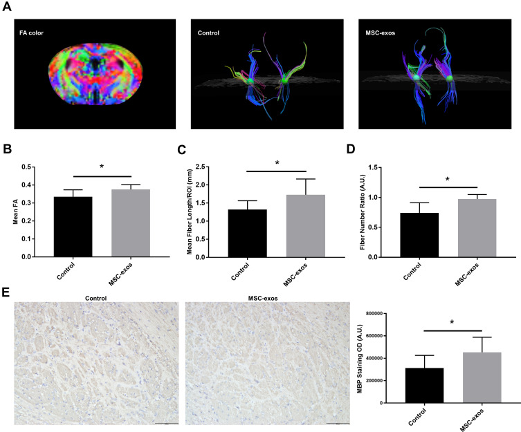
Fluorescence signals from the MSC-exos appeared in the injured brain from day 1 and peaked on day 3. The immunofluorescence staining of the brain samples revealed that the MSC-exos were localized in neurons. The behavioral scores and T2-weighted imaging indicated that the MSC-exos improved neurological functional recovery after stroke. In addition, the in vivo MR-diffusion tensor imaging (DTI) indicated that the exogenous MSC-exos increased the fractional anisotropy (FA) value, fiber length, and fiber number ratio. Furthermore, in the mice with ischemic stroke treated with MSC-exos, angiogenesis and neurogenesis were significantly improved, and the expression of IL-1β was reduced.
MSC-exos can migrate into the brains of mice with ischemic stroke and exert therapeutic effects against ischemic stroke; therefore, MSC-exos may have broad clinical applications in the future.
间充质干细胞衍生的外泌体(MSC-exos)被认为是缺血性中风的一种重要修复治疗方法。然而,外源性 MSC-exos 的迁移能力和存活率仍不清楚。在这里,我们研究了 MSC-exos 是否能迁移到缺血性大脑中,并对缺血性中风发挥保护作用。
将 DiR 标记的 MSC-exos 静脉注射到缺血性中风的小鼠体内。在第 0、1、3、5、7、10 和 14 天获取近红外荧光(NIRF)图像,并在第 1、7 和 14 天获取磁共振(MR)图像。在第 14 天,评估功能结果、血管生成、神经发生和白质重塑,并进行 Western blot 分析。
从第 1 天开始,MSC-exos 的荧光信号出现在受损的大脑中,并在第 3 天达到峰值。脑样本的免疫荧光染色显示 MSC-exos 定位于神经元中。行为评分和 T2 加权成像表明,MSC-exos 改善了中风后的神经功能恢复。此外,体内磁共振弥散张量成像(DTI)表明,外源性 MSC-exos 增加了分数各向异性(FA)值、纤维长度和纤维数量比。此外,在接受 MSC-exos 治疗的缺血性中风小鼠中,血管生成和神经发生明显改善,IL-1β 的表达减少。
MSC-exos 可以迁移到缺血性中风小鼠的大脑中,并对缺血性中风发挥治疗作用;因此,MSC-exos 在未来可能具有广泛的临床应用前景。