Department of Ophthalmology III, Centre Hospitalier National d'Ophtalmologie des Quinze-Vingts, IHU Foresight, Paris, France.
Centre Hospitalier National d'Ophtalmologie des Quinze-Vingts, INSERM-DGOS CIC 1423, IHU Foresight, Paris, France.
PLoS One. 2020 Nov 25;15(11):e0239124. doi: 10.1371/journal.pone.0239124. eCollection 2020.
To investigate the corneal epithelial thickness topography with optical coherence tomography (OCT) and its relationship with vision quality in epithelial basement membrane dystrophy (EBMD).
45 eyes of EBMD patients, 26 eyes of dry eye (DED) patients and 22 eyes of normal subjects were enrolled. All participants were subjected to 9-mm corneal epithelial mapping with OCT and vision quality was assessed with the optical quality analysis system using the objective scatter index (OSI). Central, superior, inferior, minimum, maximum, and standard deviation of epithelium thickness (Irregularity), were analysed and correlations with the OSI were calculated.
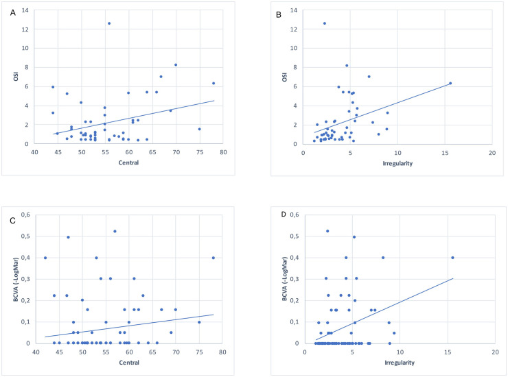
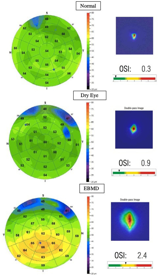
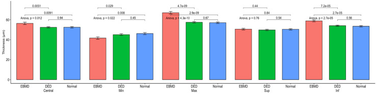
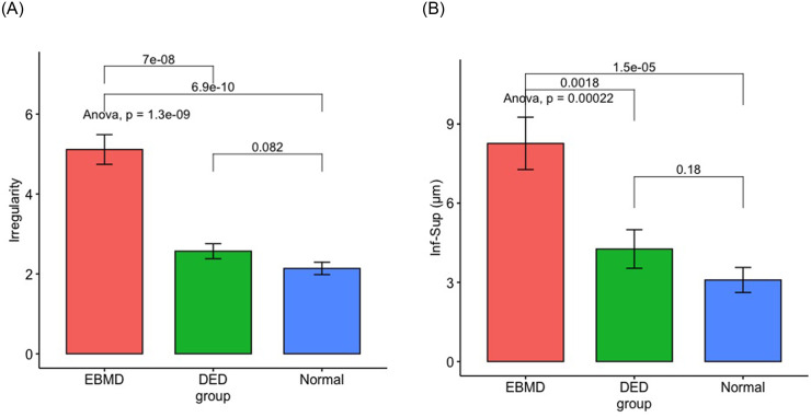
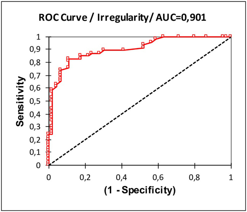
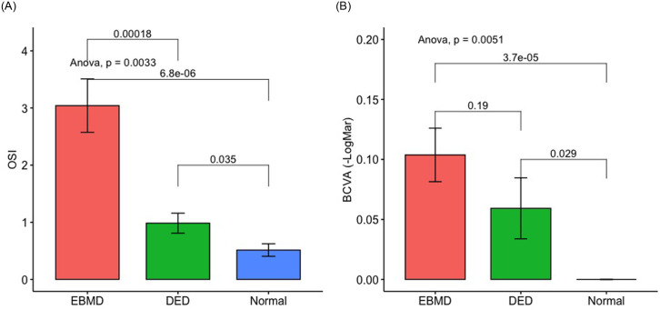
The mean (±SD) central, inferior and maximum epithelial thicknesses of the EBMD patients (respectively, 56.4 (±8.1) μm, 58.9 (±6.4) μm, and 67.1 (±8.3) μm) were thicker compared to DED patients (P<0.05) and normal subjects (P<0.05). We found greater irregularity of epithelial thickness in EBMD (5.1±2.5 μm) compared to DED patients (2.6±1.0 μm) (P = 4.4.10-6) and normal subjects (2.1±0.7 μm) (P = 7.6.10-7). The mean OSI was worse in EBMD patients than in DED patients (P = 0.01) and compared to normal subjects (P = 0.02). The OSI correlated with the epithelial thickness irregularity (Spearman coefficient = 0.54; P = 2.65.10-5).
The OCT pachymetry map demonstrated that EBMD patients had thicker corneal epithelium in the central and inferior region. These changes were correlated with objective measurements of vision quality. This OCT characterisation of the EMBD provides a better understanding of the epithelial behaviour in this dystrophy and its role in vision quality.
利用光学相干断层扫描(OCT)研究角膜上皮厚度地形图及其与上皮基底膜营养不良(EBMD)患者视力质量的关系。
纳入 45 例 EBMD 患者、26 例干眼(DED)患者和 22 例正常对照者的 9mm 角膜上皮测绘图。使用客观散射指数(OSI),通过光学生物测量分析系统评估所有参与者的视力质量。分析上皮厚度的中心、上、下、最小、最大和标准偏差(不规则性),并计算与 OSI 的相关性。
EBMD 患者的平均(±SD)中央、下方和最大上皮厚度(分别为 56.4(±8.1)μm、58.9(±6.4)μm 和 67.1(±8.3)μm)高于 DED 患者(P<0.05)和正常对照组(P<0.05)。我们发现 EBMD 患者的上皮厚度不规则性更大(5.1±2.5μm),高于 DED 患者(2.6±1.0μm)(P=4.4.10-6)和正常对照组(2.1±0.7μm)(P=7.6.10-7)。EBMD 患者的平均 OSI 比 DED 患者差(P=0.01),也比正常对照组差(P=0.02)。OSI 与上皮厚度不规则性呈正相关(Spearman 系数=0.54;P=2.65.10-5)。
OCT 角膜厚度图显示,EBMD 患者的中央和下方角膜上皮更厚。这些变化与视力质量的客观测量结果相关。该 OCT 对 EMBD 的特征描述,使我们对该营养不良中上皮的行为及其在视力质量中的作用有了更好的了解。