Tan Lan-Lan, Jiang Xiao-Lu, Xu Li-Xiao, Li Gen, Feng Chen-Xi, Ding Xin, Sun Bin, Qin Zheng-Hong, Zhang Zu-Bin, Feng Xing, Li Mei
Department of Neonatology, Children's Hospital of Soochow University, Suzhou, Jiangsu Province, China.
Department of Pediatrics Research Institute, Children's Hospital of Soochow University, Suzhou, Jiangsu Province, China.
Neural Regen Res. 2021 Jun;16(6):1037-1043. doi: 10.4103/1673-5374.300453.
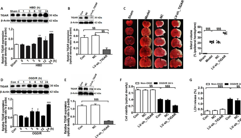
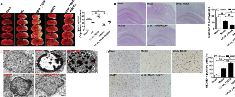
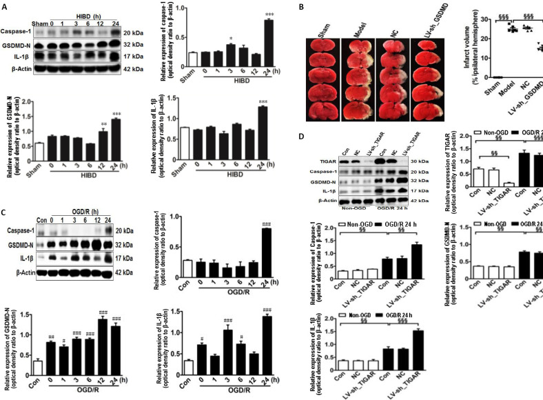
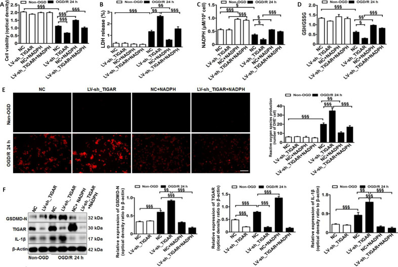
Our previous studies have demonstrated that TP53-induced glycolysis and apoptosis regulator (TIGAR) can protect neurons after cerebral ischemia/reperfusion. However, the role of TIGAR in neonatal hypoxic-ischemic brain damage (HIBD) remains unknown. In the present study, 7-day-old Sprague-Dawley rat models of HIBD were established by permanent occlusion of the left common carotid artery followed by 2-hour hypoxia. At 6 days before induction of HIBD, a lentiviral vector containing short hairpin RNA of either TIGAR or gasdermin D (LV-sh_TIGAR or LV-sh_GSDMD) was injected into the left lateral ventricle and striatum. Highly aggressively proliferating immortalized (HAPI) microglial cell models of in vitro HIBD were established by 2-hour oxygen/glucose deprivation followed by 24-hour reoxygenation. Three days before in vitro HIBD induction, HAPI microglial cells were transfected with LV-sh_TIGAR or LV-sh_GSDMD. Our results showed that TIGAR expression was increased in the neonatal rat cortex after HIBD and in HAPI microglial cells after oxygen/glucose deprivation/reoxygenation. Lentivirus-mediated TIGAR knockdown in rats markedly worsened pyroptosis and brain damage after hypoxia/ischemia in vivo and in vitro. Application of exogenous nicotinamide adenine dinucleotide phosphate (NADPH) increased the NADPH level and the glutathione/oxidized glutathione ratio and decreased reactive oxygen species levels in HAPI microglial cells after oxygen/glucose deprivation/reoxygenation. Additionally, exogenous NADPH blocked the effects of TIGAR knockdown in neonatal HIBD in vivo and in vitro. These findings show that TIGAR can inhibit microglial pyroptosis and play a protective role in neonatal HIBD. The study was approved by the Animal Ethics Committee of Soochow University of China (approval No. 2017LW003) in 2017.
我们之前的研究表明,TP53诱导的糖酵解和凋亡调节因子(TIGAR)可在脑缺血/再灌注后保护神经元。然而,TIGAR在新生儿缺氧缺血性脑损伤(HIBD)中的作用尚不清楚。在本研究中,通过永久性结扎左颈总动脉并随后进行2小时缺氧,建立了7日龄Sprague-Dawley大鼠HIBD模型。在诱导HIBD前6天,将含有TIGAR或gasdermin D短发夹RNA的慢病毒载体(LV-sh_TIGAR或LV-sh_GSDMD)注入左侧脑室和纹状体。通过2小时氧/葡萄糖剥夺并随后进行24小时复氧,建立了体外HIBD的高度侵袭性增殖永生化(HAPI)小胶质细胞模型。在体外HIBD诱导前3天,用LV-sh_TIGAR或LV-sh_GSDMD转染HAPI小胶质细胞。我们的结果表明,HIBD后新生大鼠皮质以及氧/葡萄糖剥夺/复氧后的HAPI小胶质细胞中TIGAR表达增加。慢病毒介导的大鼠TIGAR基因敲低在体内和体外均显著加重了缺氧/缺血后的细胞焦亡和脑损伤。在氧/葡萄糖剥夺/复氧后,应用外源性烟酰胺腺嘌呤二核苷酸磷酸(NADPH)可提高HAPI小胶质细胞中的NADPH水平和谷胱甘肽/氧化型谷胱甘肽比率,并降低活性氧水平。此外,外源性NADPH在体内和体外均可阻断TIGAR基因敲低对新生儿HIBD的影响。这些发现表明,TIGAR可抑制小胶质细胞焦亡,并在新生儿HIBD中发挥保护作用。该研究于2017年获得中国苏州大学动物伦理委员会批准(批准号:2017LW003)。