Clinical Molecular Diagnostic Laboratory, The Second Affiliated Hospital of Nanjing Medical University, Nanjing, Jiangsu 210003, China.
The Second Clinical Medical School of Nanjing Medical University, Nanjing, Jiangsu 210011, China.
Oxid Med Cell Longev. 2021 Nov 2;2021:6219715. doi: 10.1155/2021/6219715. eCollection 2021.
Mesenchymal stem cell-derived exosomes (MSC-exos) have been recognized as a promising therapeutic strategy for neonatal hypoxic-ischemic brain damage (HIBD). Recently, microglial pyroptosis was shown to play a vital role in the progression of neonatal HIBD. However, whether MSC-exos improve HIBD by regulating microglial pyroptosis remains unknown.
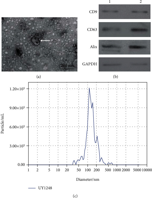
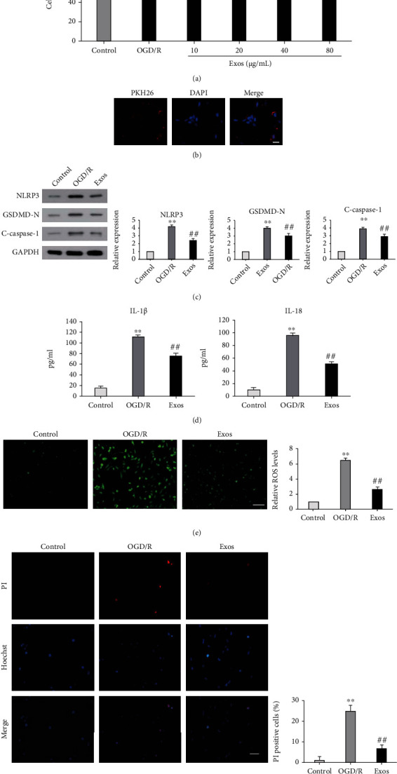
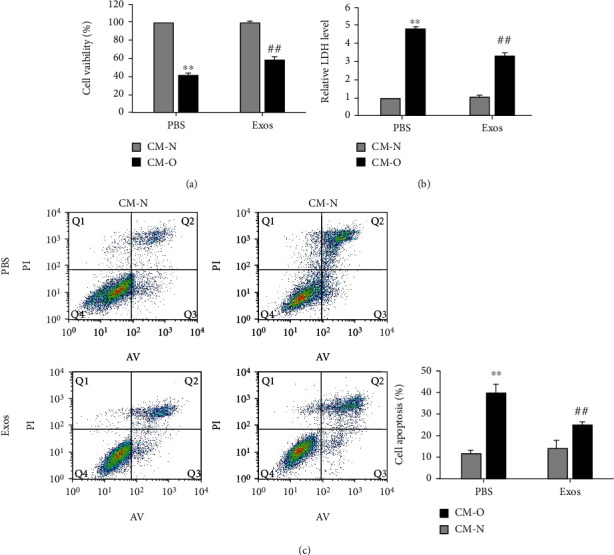
Exosomes were isolated from human umbilical cord mesenchymal stem cells (huMSCs) and identified by transmission electron microscopy (TEM), western blot, and nanoparticle tracking analysis (NTA). BV-2 cells were subjected to oxygen-glucose deprivation/reoxygenation (OGD/R) to induce microglial ischemia/reperfusion (I/R) . CCK-8, ELISA, western blot, and Hoechst 33342/PI double staining were performed to detect the pyroptosis of BV-2 cells. Conditioned medium (CM) from BV-2 cells exposed to different treatments was used to investigate its effect on neuronal injury. Moreover, 3-methyladenine (3-MA) and mitochondrial division inhibitor-1 (mdi-1) were used to verify the involvement of mitophagy in the protection of MSC-exos against OGD/R-induced pyroptosis in BV-2 cells. Finally, FOXO3a siRNA was used to investigate the involvement of FOXO3a in MSC-exo-induced mitophagy and pyroptosis inhibition.
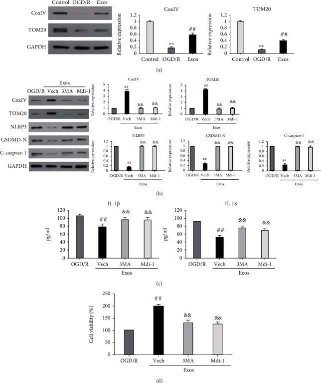
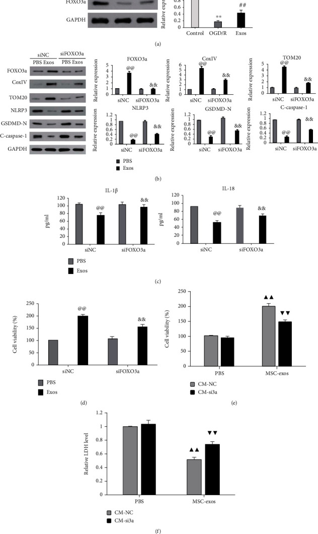
Exosomes from huMSCs were successfully extracted. In OGD/R-exposed BV-2 cells, MSC-exos increased cell viability and decreased the expression of NLRP3, cleaved caspase-1, and GSDMD-N as well as the release of IL-1 and IL-18. Compared with CM from OGD/R-exposed BV-2 cells treated with PBS, CM from OGD/R-exposed BV-2 cells treated with MSC-exos significantly increased the viability of SH-SY5Y cells and decreased LDH release. MSC-exos also increased the expression of TOM20 and COX IV in OGD/R-exposed BV-2 cells. Additionally, 3-MA and mdi-1 attenuated the inhibition of pyroptosis with MSC-exo treatment. Furthermore, FOXO3a siRNA partially abolished the neuroprotective effect of MSC-exos and attenuated mitophagy and pyroptosis inhibition induced by MSC-exo treatment.
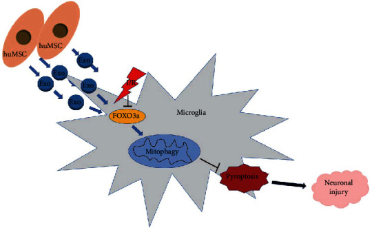
Our findings demonstrated that MSC-exos increased FOXO3a expression to enhance mitophagy, therefore protecting microglia from I/R-induced pyroptosis and alleviating subsequent neuronal injury.
间充质干细胞衍生的外泌体(MSC-exos)已被认为是治疗新生儿缺氧缺血性脑损伤(HIBD)的有前途的治疗策略。最近,小胶质细胞焦亡被证明在新生儿 HIBD 的进展中起着至关重要的作用。然而,MSC-exos 是否通过调节小胶质细胞焦亡来改善 HIBD 尚不清楚。
从人脐带间充质干细胞(huMSCs)中分离出外泌体,并通过透射电子显微镜(TEM)、western blot 和纳米颗粒跟踪分析(NTA)进行鉴定。用氧葡萄糖剥夺/复氧(OGD/R)诱导 BV-2 细胞发生小胶质细胞缺血/再灌注(I/R)。通过 CCK-8、ELISA、western blot 和 Hoechst 33342/PI 双重染色检测 BV-2 细胞的焦亡。用暴露于不同处理的 BV-2 细胞的条件培养基(CM)来研究其对神经元损伤的影响。此外,用 3-甲基腺嘌呤(3-MA)和线粒体分裂抑制剂-1(mdi-1)验证 MSC-exos 对 OGD/R 诱导的 BV-2 细胞焦亡的保护作用是否涉及自噬。最后,用 FOXO3a siRNA 研究 MSC-exo 诱导的自噬和焦亡抑制是否涉及 FOXO3a。
成功提取了 huMSCs 的外泌体。在 OGD/R 暴露的 BV-2 细胞中,MSC-exos 增加了细胞活力,降低了 NLRP3、cleaved caspase-1 和 GSDMD-N 的表达以及 IL-1 和 IL-18 的释放。与用 PBS 处理 OGD/R 暴露的 BV-2 细胞的 CM 相比,用 MSC-exos 处理 OGD/R 暴露的 BV-2 细胞的 CM 显著增加了 SH-SY5Y 细胞的活力并降低了 LDH 释放。MSC-exos 还增加了 OGD/R 暴露的 BV-2 细胞中 TOM20 和 COX IV 的表达。此外,3-MA 和 mdi-1 减弱了 MSC-exo 处理对焦亡的抑制作用。此外,FOXO3a siRNA 部分消除了 MSC-exos 的神经保护作用,并减弱了 MSC-exo 处理诱导的自噬和焦亡抑制。
我们的研究结果表明,MSC-exos 通过增加 FOXO3a 的表达来增强自噬,从而保护小胶质细胞免受 I/R 诱导的焦亡,并减轻随后的神经元损伤。