Killaars Renee C, Preuβ Myriam L G, de Vos Nathalie J P, van Berlo Camille C J L Y, Lobbes Marc B I, van der Hulst René R W J, Piatkowski Andrzej A
Department of Plastic and Reconstructive Surgery, Maastricht University Medical Centre+, Maastricht, the Netherlands.
Department of Radiology and Nuclear Medicine, Maastricht University Medical Centre+, Maastricht, the Netherlands.
Plast Reconstr Surg Glob Open. 2020 Nov 25;8(11):e3236. doi: 10.1097/GOX.0000000000003236. eCollection 2020 Nov.
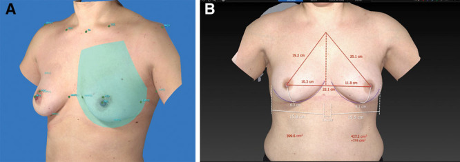
Three-dimensional (3D) camera systems are increasingly used for computerized volume calculations. In this study we investigate whether the Vectra XT 3D imaging system is a reliable tool for determination of breast volume in clinical practice. It is compared with the current gold standard in literature, magnetic resonance imaging (MRI), and current clinical practice (plastic surgeon's clinical estimation).
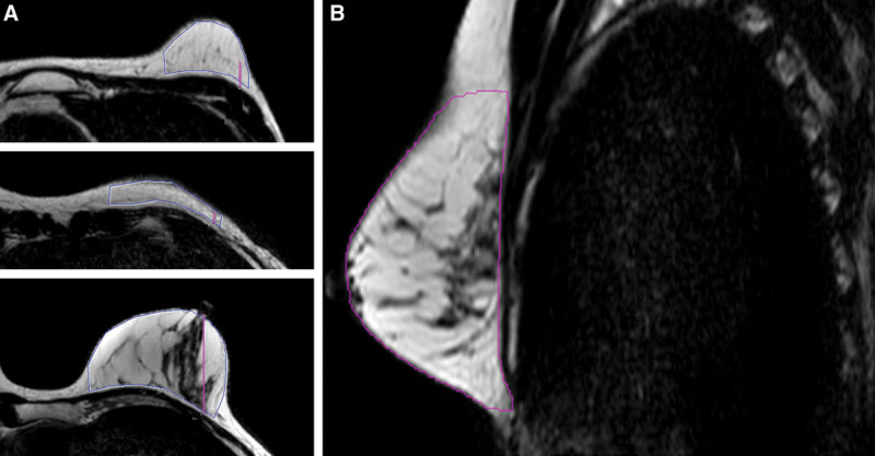
Breast volumes of 29 patients (53 breasts) were evaluated. 3D images were acquired by Vectra XT 3D imaging system. Pre-existing breast MRI images were collected. Both imaging techniques were used for volume analyses, calculated by two independent investigators. Breast volume estimations were done by plastic surgeons during outpatient consultations. All volume measurements were compared using paired samples -test, intra-class correlation coefficient, Pearson's correlation, and Bland-Altman analysis.
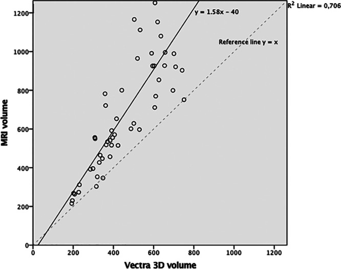
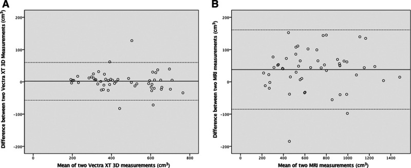
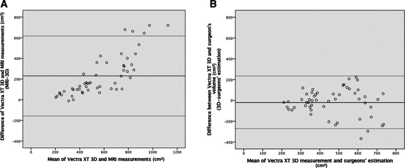
Two 3D breast volume measurements showed an excellent reliability (intra-class correlation coefficient: 0.991), which was comparable to the reliability of MRI measurements (intra-class correlation coefficient: 0.990). Mean (SD) breast volume measured with 3D breast volume was 454 cm (157) and with MRI was 687 cm (312). These volumes were significantly different, but a linear association could be found: y(MRI) = 1.58 × (3D) - 40. Three-dimensional breast volume was not significantly different from volume estimation made by plastic surgeons (472 cm (69), = 0.323).
The 3D imaging system measures lower volumes for breasts than MRI. However, 3D measurements show a linear association with MRI and have excellent reliability, making them an objective and reproducible measuring method suitable for clinical practice.
三维(3D)摄像系统越来越多地用于计算机化容积计算。在本研究中,我们调查了Vectra XT 3D成像系统在临床实践中用于确定乳房容积是否是一种可靠的工具。将其与文献中当前的金标准磁共振成像(MRI)以及当前临床实践(整形外科医生的临床估计)进行比较。
评估了29例患者(53个乳房)的乳房容积。通过Vectra XT 3D成像系统获取3D图像。收集现有的乳房MRI图像。两种成像技术均用于容积分析,由两名独立研究人员进行计算。乳房容积估计由整形外科医生在门诊会诊时完成。使用配对样本t检验、组内相关系数、Pearson相关性和Bland-Altman分析对所有容积测量值进行比较。
两次3D乳房容积测量显示出极佳的可靠性(组内相关系数:0.991),这与MRI测量的可靠性相当(组内相关系数:0.990)。3D乳房容积测量的平均(标准差)乳房容积为454 cm³(157),MRI测量的为687 cm³(312)。这些容积有显著差异,但可以发现线性关联:y(MRI)= 1.58 × (3D) - 40。三维乳房容积与整形外科医生的容积估计(472 cm³(69),P = 0.323)无显著差异。
3D成像系统测量的乳房容积低于MRI。然而,3D测量与MRI显示出线性关联且具有极佳的可靠性,使其成为一种适用于临床实践的客观且可重复的测量方法。