Chen Keying, Wellman Steven M, Yaxiaer Yalikun, Eles James R, Kozai Takashi Dy
Department of Bioengineering, University of Pittsburgh, USA; Center for the Neural Basis of Cognition, University of Pittsburgh and Carnegie Mellon University, USA.
Eberly College of Science, Pennsylvania State University, University Park, USA.
Biomaterials. 2021 Jan;268:120526. doi: 10.1016/j.biomaterials.2020.120526. Epub 2020 Nov 23.
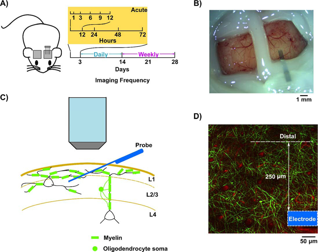
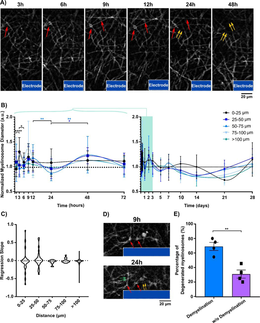
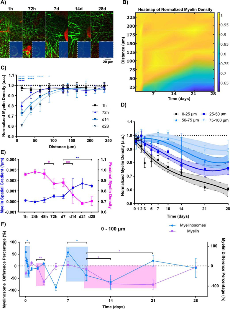
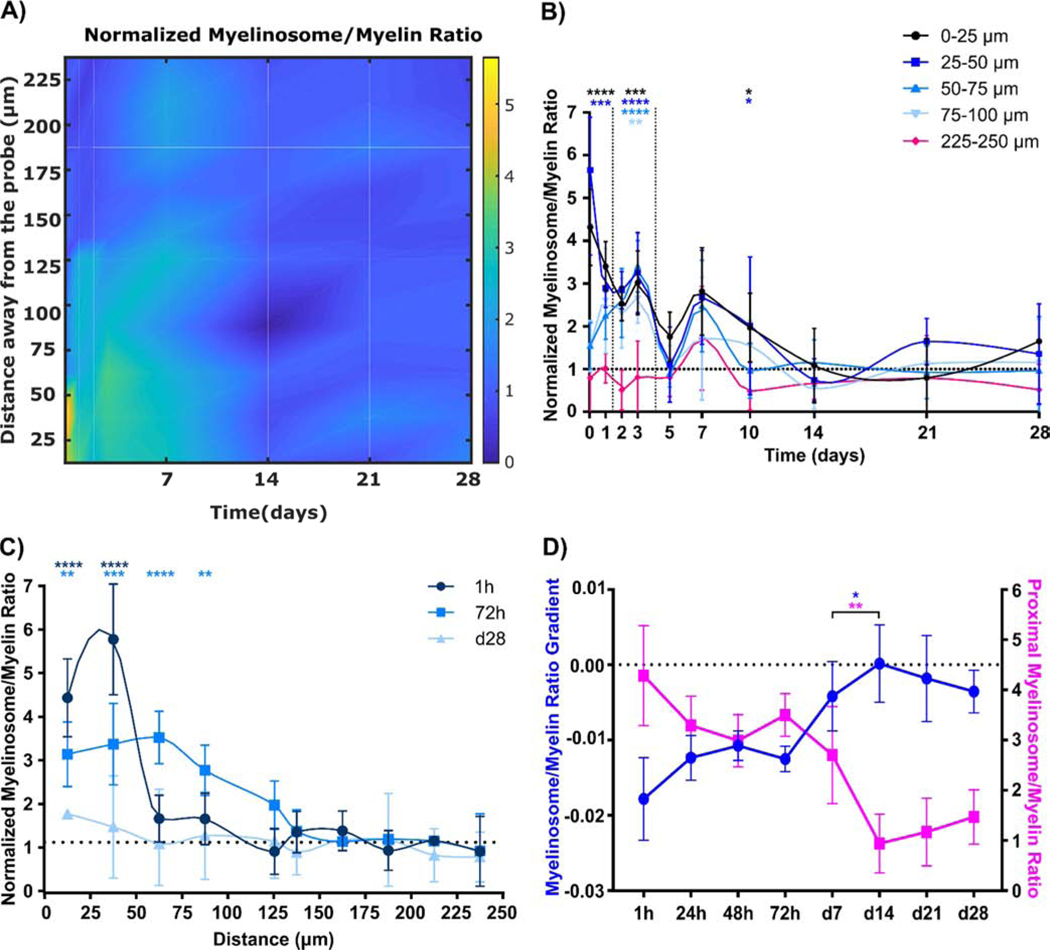
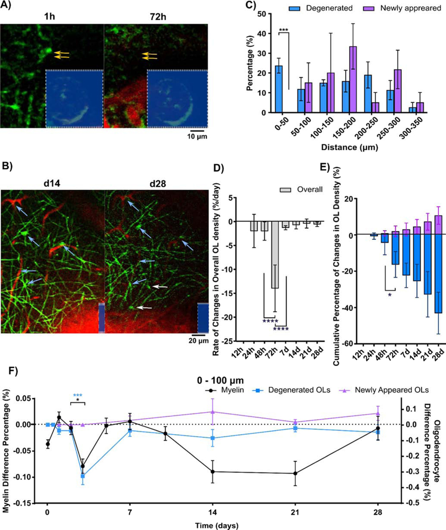
Intracortical microelectrodes with the ability to detect intrinsic electrical signals and/or deliver electrical stimulation into local brain regions have been a powerful tool to understand brain circuitry and for therapeutic applications to neurological disorders. However, the chronic stability and sensitivity of these intracortical microelectrodes are challenged by overwhelming biological responses, including severe neuronal loss and thick glial encapsulation. Unlike microglia and astrocytes whose activity have been extensively examined, oligodendrocytes and their myelin processes remain poorly studied within the neural interface field. Oligodendrocytes have been widely recognized to modulate electrical signal conductance along axons through insulating myelin segments. Emerging evidence offers an alternative perspective on neuron-oligodendrocyte coupling where oligodendrocytes provide metabolic and neurotrophic support to neurons through cytoplasmic myelin channels and monocarboxylate transporters. This study uses in vivo multi-photon microscopy to gain insights into the dynamics of oligodendrocyte soma and myelin processes in response to chronic device implantation injury over 4 weeks. We observe that implantation induces acute oligodendrocyte injury including initial deformation and substantial myelinosome formation, an early sign of myelin injury. Over chronic implantation periods, myelin and oligodendrocyte soma suffer severe degeneration proximal to the interface. Interestingly, wound healing attempts such as oligodendrogenesis are initiated over time, however they are hampered by continued degeneration near the implant. Nevertheless, this detailed characterization of oligodendrocyte spatiotemporal dynamics during microelectrode-induced inflammation may provide insights for novel intervention targets to facilitate oligodendrogenesis, enhance the integration of neural-electrode interfaces, and improve long-term functional performance.
能够检测内在电信号和/或将电刺激传递到局部脑区的皮层内微电极,一直是理解脑回路以及用于神经疾病治疗应用的有力工具。然而,这些皮层内微电极的长期稳定性和灵敏度受到压倒性生物反应的挑战,包括严重的神经元损失和厚厚的胶质细胞包裹。与小胶质细胞和星形胶质细胞的活性已被广泛研究不同,少突胶质细胞及其髓鞘过程在神经界面领域仍研究较少。少突胶质细胞已被广泛认为可通过绝缘的髓鞘节段调节沿轴突的电信号传导。新出现的证据为神经元 - 少突胶质细胞耦合提供了另一种观点,即少突胶质细胞通过细胞质髓鞘通道和单羧酸转运蛋白为神经元提供代谢和神经营养支持。本研究使用体内多光子显微镜来深入了解少突胶质细胞胞体和髓鞘过程在长达4周的慢性装置植入损伤后的动态变化。我们观察到植入会引起急性少突胶质细胞损伤,包括初始变形和大量髓鞘小体形成,这是髓鞘损伤的早期迹象。在慢性植入期间,髓鞘和少突胶质细胞胞体在界面近端会严重退化。有趣的是,随着时间的推移会启动诸如少突胶质细胞生成等伤口愈合尝试,然而它们受到植入物附近持续退化的阻碍。尽管如此,这种对微电极诱导炎症期间少突胶质细胞时空动态的详细表征,可能为促进少突胶质细胞生成、增强神经 - 电极界面整合以及改善长期功能性能的新型干预靶点提供见解。