Cognitive Science Research Group, Korea Brain Research Institute, Daegu, South Korea.
Department of Psychiatry, College of Medicine, The Catholic University of Korea, 222 Banpo-daero, Seocho-gu, Seoul, 137-701, South Korea.
BMC Psychiatry. 2020 Dec 10;20(1):586. doi: 10.1186/s12888-020-02972-8.
Electroencephalography (EEG) frontal alpha asymmetry (FAA) has been observed in several psychiatric disorders. Dominance in left or right frontal alpha activity remains inconsistent in patients with major depressive disorder (MDD), patients with schizophrenia, and healthy controls. This study compared FAA among patients with MDD and schizophrenia, and healthy controls.
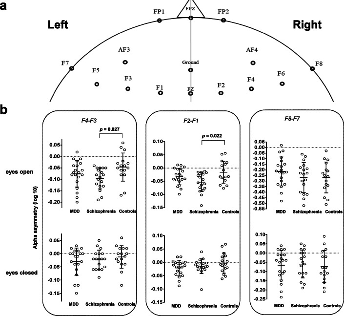
We recruited 20 patients with MDD, 18 patients with schizophrenia, and 16 healthy individuals. The EEG alpha frequency ranged from 8 Hz to 12 Hz. FAA was expressed as the difference between absolute power values of right and left hemisphere electrodes in the alpha frequency range (common-log-transformed frontal right- and left-hemisphere electrodes: F4-F3, F8-F7, FP2-FP1, AF4-AF3, F6-F5, and F2-F1). Hamilton depression and anxiety rating scales were evaluated in patients with MDD. Positive and negative syndrome scales were evaluated in patients with schizophrenia.
Patients with schizophrenia showed significantly lower left FAA than healthy controls (F4-F3, schizophrenia vs. healthy controls: - 0.10 ± 0.04 vs. -0.05 ± 0.05). There were no significant differences in FAA between patients with schizophrenia and MDD as well as between patients with MDD and healthy controls.
The present study suggests that FAA indicates a relatively lower activation of left frontal electrodes in schizophrenia. The left-lateralized FAA could be a neuropathological attribute in patients with schizophrenia, but a lack of sample size and information such as medication and duration of illness might obscure the interpretation and generalization of our findings. Thus, further studies to verify the findings would be warranted.
脑电图(EEG)额区阿尔法不对称(FAA)在几种精神疾病中都有观察到。在重度抑郁症(MDD)患者、精神分裂症患者和健康对照者中,左或右额区阿尔法活动优势并不一致。本研究比较了 MDD 患者、精神分裂症患者和健康对照者的 FAA。
我们招募了 20 名 MDD 患者、18 名精神分裂症患者和 16 名健康个体。EEG 阿尔法频率范围为 8-12Hz。FAA 表示左右半球电极在阿尔法频带(共对数转换的额区右左半球电极:F4-F3、F8-F7、FP2-FP1、AF4-AF3、F6-F5 和 F2-F1)之间的绝对功率值的差异。MDD 患者接受汉密尔顿抑郁和焦虑评定量表评估,精神分裂症患者接受阳性和阴性综合征量表评估。
精神分裂症患者的左 FAA 显著低于健康对照组(F4-F3,精神分裂症 vs 健康对照组:-0.10±0.04 对 -0.05±0.05)。精神分裂症患者与 MDD 患者以及 MDD 患者与健康对照组之间的 FAA 没有显著差异。
本研究表明 FAA 表明精神分裂症患者左额电极的激活相对较低。左侧 FAA 可能是精神分裂症患者的一种神经病理学特征,但由于样本量较小以及用药和疾病持续时间等信息缺失,可能会使我们的研究结果的解释和推广变得模糊。因此,有必要进行进一步的研究来验证这些发现。