• 文献检索
  • 文档翻译
  • 深度研究
  • 学术资讯
  • Suppr Zotero 插件Zotero 插件
  • 邀请有礼
  • 套餐&价格
  • 历史记录
应用&插件
Suppr Zotero 插件Zotero 插件浏览器插件Mac 客户端Windows 客户端微信小程序
定价
高级版会员购买积分包购买API积分包
服务
文献检索文档翻译深度研究API 文档MCP 服务
关于我们
关于 Suppr公司介绍联系我们用户协议隐私条款
关注我们

Suppr 超能文献

核心技术专利:CN118964589B侵权必究
粤ICP备2023148730 号-1Suppr @ 2026

文献检索

告别复杂PubMed语法,用中文像聊天一样搜索,搜遍4000万医学文献。AI智能推荐,让科研检索更轻松。

立即免费搜索

文件翻译

保留排版,准确专业,支持PDF/Word/PPT等文件格式,支持 12+语言互译。

免费翻译文档

深度研究

AI帮你快速写综述,25分钟生成高质量综述,智能提取关键信息,辅助科研写作。

立即免费体验

前列腺癌患者同时进行[镓]镓-PSMA-11 PET/MRI成像时SUVmax的病变与背景比值阈值

Lesion-to-background ratio threshold value of SUVmax of simultaneous [Ga]Ga-PSMA-11 PET/MRI imaging in patients with prostate cancer.

作者信息

Zhao Jing, Hamm Bernd, Brenner Winfried, Makowski Marcus R

机构信息

Institute of Radiology and Nuclear Medicine, Charité - Universitätsmedizin Berlin, corporate member of Freie Universität Berlin, Humboldt-Universität zu Berlin, and Berlin Institute of Health, Charitéplatz 1, 10117, Berlin, Germany.

Institute of Nuclear Medicine, Charité - Universitätsmedizin Berlin, corporate member of Freie Universität Berlin, Humboldt-Universität zu Berlin, and Berlin Institute of Health, Augustenburger Platz 1, 13353, Berlin, Germany.

出版信息

Insights Imaging. 2020 Dec 17;11(1):137. doi: 10.1186/s13244-020-00926-y.

DOI:10.1186/s13244-020-00926-y
PMID:33331959
原文链接:https://pmc.ncbi.nlm.nih.gov/articles/PMC7745090/
Abstract

PURPOSE

This study aimed to calculate an applicable relative ratio threshold value instead of the absolute threshold value for simultaneous Ga prostate-specific membrane antigen/positron emission tomography ([Ga]Ga-PSMA-11 PET) in patients with prostate cancer (PCa).

MATERIALS AND METHODS

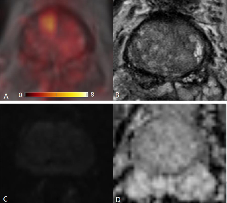
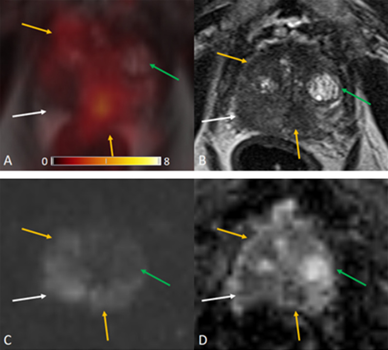
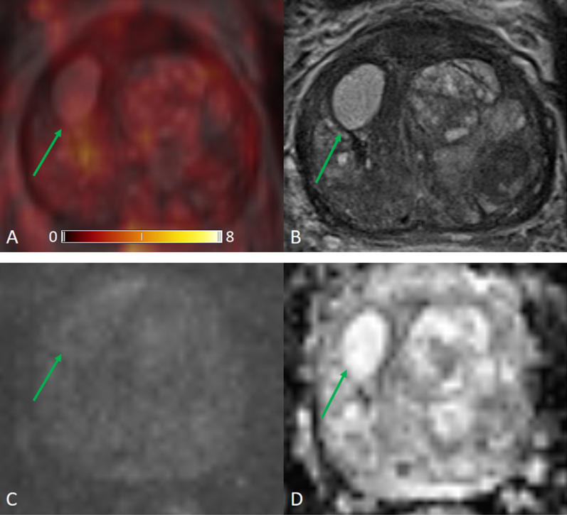
Our study evaluated thirty-two patients and 170 focal prostate lesions. Lesions are classified into groups according to Prostate Imaging Reporting and Data System (PI-RADS). Standardized uptake values maximum (SUVmax), corresponding lesion-to-background ratios (LBRs) of SUVmax, and LBR distributions of each group were measured based on regions of interest (ROI). We examined LBR with receiver operating characteristic analysis to determine threshold values for differentiation between multiparametric magnetic resonance imaging (mpMRI)-positive and mpMRI-negative lesions.

RESULTS

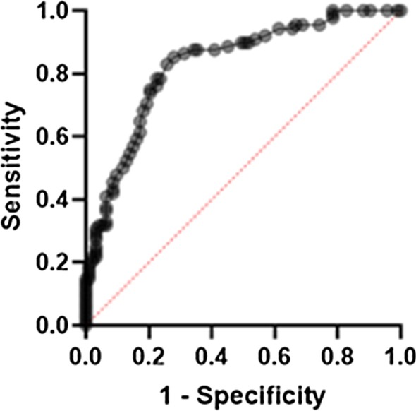
We analyzed a total of 170 focal prostate lesions. Lesions number of PI-RADS 2 to 5 was 70, 16, 46, and 38. LBR of SUVmax of each PI-RADS scores was 1.5 (0.9, 2.4), 2.5 (1.6, 3.4), 3.7 (2.6, 4.8), and 6.7 (3.5, 12.7). Based on an optimal threshold ratio of 2.5 to be exceeded, lesions could be classified into MRI-positive lesion on [Ga]Ga-PSMA PET with a sensitivity of 85.2%, a specificity of 72.0%, with the corresponding area under the receiver operating characteristic curve (AUC) of 0.83, p < 0.001. This value matches the imaging findings better.

CONCLUSION

The ratio threshold value of SUVmax, LBR, has improved clinical and research applicability compared with the absolute value of SUVmax. A higher threshold value than the background's uptake can dovetail the imaging findings on MRI better. It reduces the bias from using absolute background uptake value as the threshold value.

摘要

目的

本研究旨在计算一个适用于前列腺癌(PCa)患者的相对比值阈值,以替代同时进行镓前列腺特异性膜抗原/正电子发射断层扫描([Ga]Ga-PSMA-11 PET)时的绝对阈值。

材料与方法

我们的研究评估了32例患者和170个前列腺局灶性病变。病变根据前列腺影像报告和数据系统(PI-RADS)进行分组。基于感兴趣区域(ROI)测量标准化摄取值最大值(SUVmax)、SUVmax相应的病变与背景比值(LBR)以及每组的LBR分布。我们通过接受者操作特征分析检查LBR,以确定多参数磁共振成像(mpMRI)阳性和mpMRI阴性病变之间的鉴别阈值。

结果

我们共分析了170个前列腺局灶性病变。PI-RADS 2至5级的病变数量分别为70、16、46和38个。每个PI-RADS评分的SUVmax的LBR分别为1.5(0.9,2.4)、2.5(1.6,3.4)、3.7(2.6,4.8)和6.7(3.5,12.7)。基于超过2.5的最佳阈值比值,病变在[Ga]Ga-PSMA PET上可被分类为MRI阳性病变,灵敏度为85.2%,特异性为72.0%,相应的接受者操作特征曲线下面积(AUC)为0.83,p < 0.001。该值与影像学表现更匹配。

结论

与SUVmax的绝对值相比,SUVmax的比值阈值LBR提高了临床和研究适用性。高于背景摄取的阈值能更好地与MRI上的影像学表现相吻合。它减少了将绝对背景摄取值用作阈值所带来的偏差。

https://cdn.ncbi.nlm.nih.gov/pmc/blobs/553f/7745090/966e23e4229a/13244_2020_926_Fig6_HTML.jpg
https://cdn.ncbi.nlm.nih.gov/pmc/blobs/553f/7745090/1a2f4ca789c9/13244_2020_926_Fig1_HTML.jpg
https://cdn.ncbi.nlm.nih.gov/pmc/blobs/553f/7745090/28648d838571/13244_2020_926_Fig2_HTML.jpg
https://cdn.ncbi.nlm.nih.gov/pmc/blobs/553f/7745090/f7cea32a7867/13244_2020_926_Fig3_HTML.jpg
https://cdn.ncbi.nlm.nih.gov/pmc/blobs/553f/7745090/976e63e6a694/13244_2020_926_Fig4_HTML.jpg
https://cdn.ncbi.nlm.nih.gov/pmc/blobs/553f/7745090/c2f59cfe242f/13244_2020_926_Fig5_HTML.jpg
https://cdn.ncbi.nlm.nih.gov/pmc/blobs/553f/7745090/966e23e4229a/13244_2020_926_Fig6_HTML.jpg

相似文献

1
Lesion-to-background ratio threshold value of SUVmax of simultaneous [Ga]Ga-PSMA-11 PET/MRI imaging in patients with prostate cancer.前列腺癌患者同时进行[镓]镓-PSMA-11 PET/MRI成像时SUVmax的病变与背景比值阈值
Insights Imaging. 2020 Dec 17;11(1):137. doi: 10.1186/s13244-020-00926-y.
2
Correlation between Intraprostatic PSMA Uptake and MRI PI-RADS of [Ga]Ga-PSMA-11 PET/MRI in Patients with Prostate Cancer: Comparison of PI-RADS Version 2.0 and PI-RADS Version 2.1.前列腺癌患者中[镓]镓-PSMA-11 PET/MRI的前列腺内PSMA摄取与MRI PI-RADS的相关性:PI-RADS第2.0版与PI-RADS第2.1版的比较
Cancers (Basel). 2020 Nov 26;12(12):3523. doi: 10.3390/cancers12123523.
3
More advantages in detecting bone and soft tissue metastases from prostate cancer using F-PSMA PET/CT.使用F-PSMA PET/CT检测前列腺癌骨和软组织转移方面有更多优势。
Hell J Nucl Med. 2019 Jan-Apr;22(1):6-9. doi: 10.1967/s002449910952. Epub 2019 Mar 7.
4
PI-RADS-Based Segmented Threshold of PSMA-PET SUVmax Is Better than Traditional Fixed Threshold for Diagnosing Clinically Significant Prostate Cancer Especially for PI-RADS 3 Lesions.基于PI-RADS的PSMA-PET SUVmax分段阈值在诊断临床显著前列腺癌方面优于传统固定阈值,尤其对于PI-RADS 3类病变。
Mol Imaging Biol. 2023 Oct;25(5):887-896. doi: 10.1007/s11307-023-01841-6. Epub 2023 Jul 25.
5
Diagnostic value of integrated F-PSMA-1007 PET/MRI compared with that of biparametric MRI for the detection of prostate cancer.集成F-PSMA-1007 PET/MRI与双参数MRI在检测前列腺癌方面的诊断价值比较
Prostate Int. 2022 Jun;10(2):108-116. doi: 10.1016/j.prnil.2022.03.003. Epub 2022 Mar 28.
6
Diagnostic accuracy of Ga-prostate-specific membrane antigen (PSMA) positron-emission tomography (PET) and multiparametric (mp)MRI to detect intermediate-grade intra-prostatic prostate cancer using whole-mount pathology: impact of the addition of Ga-PSMA PET to mpMRI.使用全层病理检测前列腺中级前列腺癌时,镓标记前列腺特异性膜抗原(PSMA)正电子发射断层扫描(PET)和多参数(mp)MRI的诊断准确性:在mpMRI中添加镓标记PSMA PET的影响
BJU Int. 2019 Nov;124 Suppl 1:42-49. doi: 10.1111/bju.14794. Epub 2019 Jul 9.
7
Multiparametric Magnetic Resonance Imaging, Ga Prostate-Specific Membrane Antigen Positron Emission Tomography-Computed Tomography, and Respective Quantitative Parameters in Detection and Localization of Clinically Significant Prostate Cancer in Intermediate- and High-Risk Group Patients: An Indian Demographic Study.多参数磁共振成像、镓前列腺特异性膜抗原正电子发射断层扫描-计算机断层扫描以及各自的定量参数在中高危组患者临床显著性前列腺癌检测与定位中的应用:一项印度人群研究
Indian J Nucl Med. 2021 Oct-Dec;36(4):362-370. doi: 10.4103/ijnm.ijnm_80_21. Epub 2021 Dec 15.
8
The use of Ga-PSMA PET/CT to stratify patients with PI-RADS 3 lesions according to clinically significant prostate cancer risk.使用镓-前列腺特异性膜抗原正电子发射断层扫描/计算机断层扫描(Ga-PSMA PET/CT)根据具有临床意义的前列腺癌风险对前列腺影像报告和数据系统(PI-RADS)3类病变患者进行分层。
Prostate. 2023 Apr;83(5):430-439. doi: 10.1002/pros.24475. Epub 2022 Dec 21.
9
Simultaneous Ga-PSMA HBED-CC PET/MRI Improves the Localization of Primary Prostate Cancer.镓-PSMA HBED-CC PET/MRI 联合检查可提高前列腺癌原发灶的定位诊断。
Eur Urol. 2016 Nov;70(5):829-836. doi: 10.1016/j.eururo.2015.12.053. Epub 2016 Jan 18.
10
Diagnostic Performance of Ga Prostate-specific Membrane Antigen PET/MRI Compared with Multiparametric MRI for Detecting Clinically Significant Prostate Cancer.镓前列腺特异性膜抗原 PET/MRI 与多参数 MRI 检测临床显著前列腺癌的诊断性能比较。
Radiology. 2021 Nov;301(2):379-386. doi: 10.1148/radiol.2021204093. Epub 2021 Aug 31.

引用本文的文献

1
Standardized template for clinical reporting of PSMA PET/CT scans.PSMA PET/CT 扫描临床报告的标准化模板。
Eur J Nucl Med Mol Imaging. 2024 Dec;52(1):335-341. doi: 10.1007/s00259-024-06857-w. Epub 2024 Aug 15.
2
Target Volume Optimization for Localized Prostate Cancer.局限性前列腺癌的靶区优化。
Pract Radiat Oncol. 2024 Nov-Dec;14(6):522-540. doi: 10.1016/j.prro.2024.06.006. Epub 2024 Jul 15.
3
Can the Gleason score be predicted in patients with prostate cancer? A dynamic contrast-enhanced MRI, Ga-PSMA PET/CT, PSA, and PSA-density comparison study.

本文引用的文献

1
Variability of the Positive Predictive Value of PI-RADS for Prostate MRI across 26 Centers: Experience of the Society of Abdominal Radiology Prostate Cancer Disease-focused Panel.26 家中心前列腺 MRI 中 PI-RADS 的阳性预测值的可变性:腹放射学会前列腺癌疾病重点专家组的经验。
Radiology. 2020 Jul;296(1):76-84. doi: 10.1148/radiol.2020190646. Epub 2020 Apr 21.
2
Correlation of SUV and Apparent Diffusion Coefficient Values Detected by Ga-68 PSMA PET/MRI in Primary Prostate Lesions and Their Significance in Lymph Node Metastasis: Preliminary Results of an On-going Study.Ga-68 PSMA PET/MRI检测原发性前列腺病变中SUV与表观扩散系数值的相关性及其在淋巴结转移中的意义:一项正在进行的研究的初步结果
Mol Imaging Radionucl Ther. 2019 Sep 6;28(3):104-111. doi: 10.4274/mirt.galenos.2019.63825.
3
前列腺癌患者的格里森评分可以预测吗?一项动态对比增强 MRI、Ga-PSMA PET/CT、PSA 和 PSA 密度比较研究。
Diagn Interv Radiol. 2023 Sep 5;29(5):647-655. doi: 10.4274/dir.2023.232186. Epub 2023 Jul 3.
4
A Systematic Review of the Variability in Performing and Reporting Intraprostatic Prostate-specific Membrane Antigen Positron Emission Tomography in Primary Staging Studies.原发性分期研究中前列腺内前列腺特异性膜抗原正电子发射断层扫描操作与报告变异性的系统评价
Eur Urol Open Sci. 2023 Mar 3;50:91-105. doi: 10.1016/j.euros.2023.01.010. eCollection 2023 Apr.
5
Utility of Biology-Guided Radiotherapy to Metastases Diagnosed During Staging of High-Risk Biopsy-Proven Prostate Cancer.生物学引导放疗在高危活检证实前列腺癌分期时诊断出的转移灶中的应用
Front Oncol. 2022 Apr 12;12:854589. doi: 10.3389/fonc.2022.854589. eCollection 2022.
6
Establishment and prospective validation of an SUV cutoff value to discriminate clinically significant prostate cancer from benign prostate diseases in patients with suspected prostate cancer by Ga-PSMA PET/CT: a real-world study.应用 Ga-PSMA PET/CT 建立并前瞻性验证 SUV 截断值以鉴别疑似前列腺癌患者中由前列腺特异性膜抗原 PET/CT 检测的临床显著前列腺癌与良性前列腺疾病:一项真实世界研究。
Theranostics. 2021 Jul 25;11(17):8396-8411. doi: 10.7150/thno.58140. eCollection 2021.
7
Dynamic Contrast-Enhanced MRI of Prostate Lesions of Simultaneous [Ga]Ga-PSMA-11 PET/MRI: Comparison between Intraprostatic Lesions and Correlation between Perfusion Parameters.同时进行[镓]Ga-PSMA-11 PET/MRI的前列腺病变的动态对比增强MRI:前列腺内病变之间的比较及灌注参数的相关性
Cancers (Basel). 2021 Mar 19;13(6):1404. doi: 10.3390/cancers13061404.
8
Molecular MR Imaging of Prostate Cancer.前列腺癌的分子磁共振成像
Biomedicines. 2020 Dec 22;9(1):1. doi: 10.3390/biomedicines9010001.
Whole-Body Integrated [Ga]PSMA-11-PET/MR Imaging in Patients with Recurrent Prostate Cancer: Comparison with Whole-Body PET/CT as the Standard of Reference.全身整合 [Ga]PSMA-11-PET/MR 成像在复发性前列腺癌患者中的应用:与全身 PET/CT 作为参考标准的比较。
Mol Imaging Biol. 2020 Jun;22(3):788-796. doi: 10.1007/s11307-019-01424-4.
4
Comparison between pelvic PSMA-PET/MR and whole-body PSMA-PET/CT for the initial evaluation of prostate cancer: a proof of concept study.盆腔 PSMA-PET/MR 与全身 PSMA-PET/CT 用于前列腺癌初始评估的比较:一项概念验证研究。
Eur Radiol. 2020 Jan;30(1):328-336. doi: 10.1007/s00330-019-06353-y. Epub 2019 Jul 22.
5
Detection Rate of F-Labeled PSMA PET/CT in Biochemical Recurrent Prostate Cancer: A Systematic Review and a Meta-Analysis.F 标记的 PSMA PET/CT 在生化复发前列腺癌中的检测率:一项系统评价和荟萃分析
Cancers (Basel). 2019 May 23;11(5):710. doi: 10.3390/cancers11050710.
6
Ga-PSMA-11 PET/MR Detects Local Recurrence Occult on mpMRI in Prostate Cancer Patients After HIFU.镓-PSMA-11 PET/MR 检测高强度聚焦超声治疗后前列腺癌患者 mpMRI 隐匿性局部复发
J Nucl Med. 2019 Aug;60(8):1118-1123. doi: 10.2967/jnumed.118.221564. Epub 2019 Jan 25.
7
Combination of Ga-PSMA PET/CT and Multiparametric MRI Improves the Detection of Clinically Significant Prostate Cancer: A Lesion-by-Lesion Analysis.镓-PSMA PET/CT 与多参数 MRI 的联合应用提高了临床显著前列腺癌的检出率:基于病灶的分析。
J Nucl Med. 2019 Jul;60(7):944-949. doi: 10.2967/jnumed.118.221010. Epub 2018 Dec 14.
8
Metaanalysis of Ga-PSMA-11 PET Accuracy for the Detection of Prostate Cancer Validated by Histopathology.基于组织病理学验证的 Ga-PSMA-11 PET 检测前列腺癌准确性的荟萃分析
J Nucl Med. 2019 Jun;60(6):786-793. doi: 10.2967/jnumed.118.219501. Epub 2018 Dec 7.
9
Improved specificity with Ga PSMA PET/CT to detect clinically significant lesions "invisible" on multiparametric MRI of the prostate: a single institution comparative analysis with radical prostatectomy histology.与多参数前列腺 MRI 相比,Ga PSMA PET/CT 检测前列腺癌临床显著病灶的特异性提高:与根治性前列腺切除术组织学的单机构比较分析。
Eur J Nucl Med Mol Imaging. 2019 Jan;46(1):20-30. doi: 10.1007/s00259-018-4160-7. Epub 2018 Sep 19.
10
Diagnostic Accuracy of Ga-PSMA-11 PET/MRI Compared with Multiparametric MRI in the Detection of Prostate Cancer.镓-PSMA-11 PET/MRI 与多参数 MRI 检测前列腺癌的诊断准确性比较。
Radiology. 2018 Dec;289(3):730-737. doi: 10.1148/radiol.2018180788. Epub 2018 Sep 18.