Division of Cardiovascular Medicine, Department of Medicine, Center for Interdisciplinary Cardiovascular Sciences, Brigham and Women's Hospital, Harvard Medical School, Boston, Massachusetts, USA.
Division of Cardiovascular Medicine, Department of Medicine, Center for Interdisciplinary Cardiovascular Sciences, Brigham and Women's Hospital, Harvard Medical School, Boston, Massachusetts, USA; Channing Division of Network Medicine, Brigham and Women's Hospital, Harvard Medical School, Boston, Massachusetts, USA.
J Biol Chem. 2021 Jan-Jun;296:100193. doi: 10.1074/jbc.RA120.015700. Epub 2021 Jan 6.
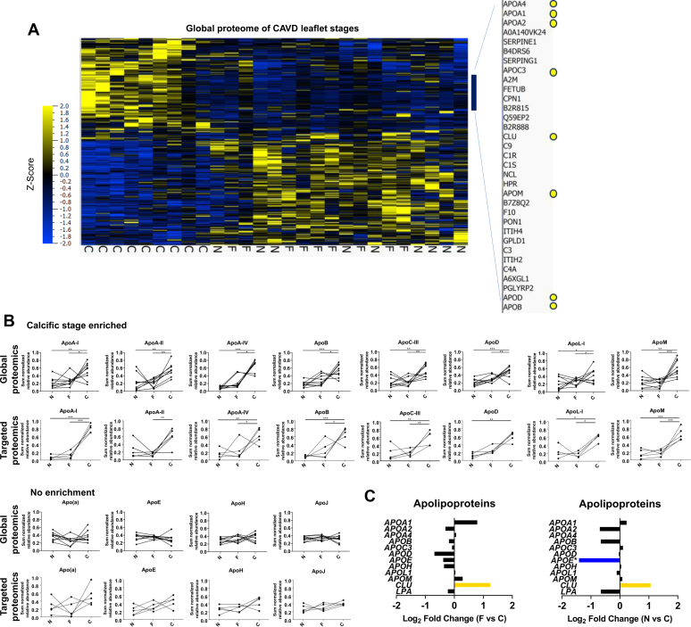
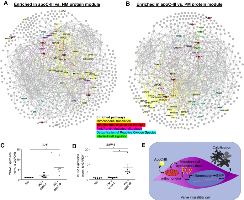
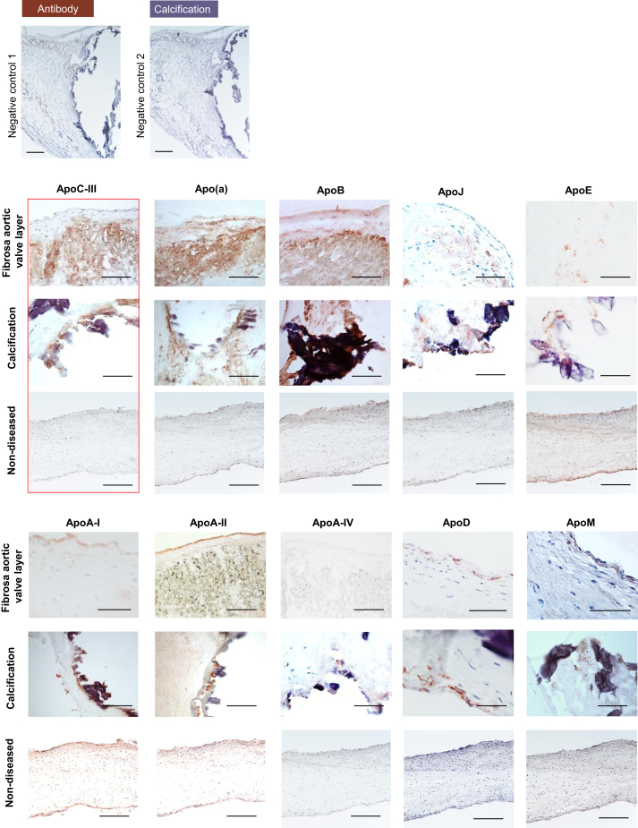
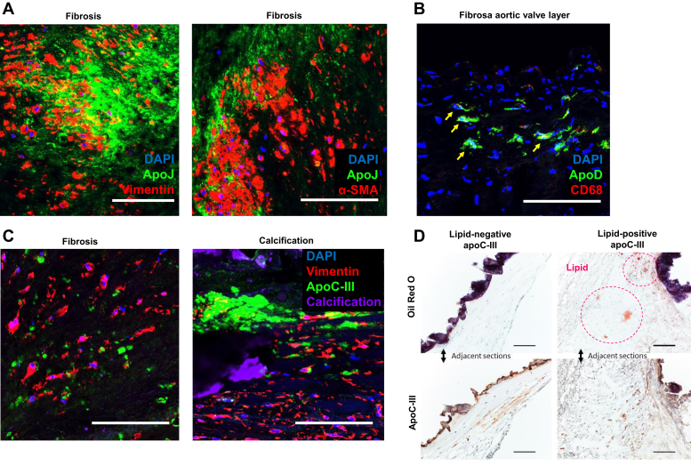
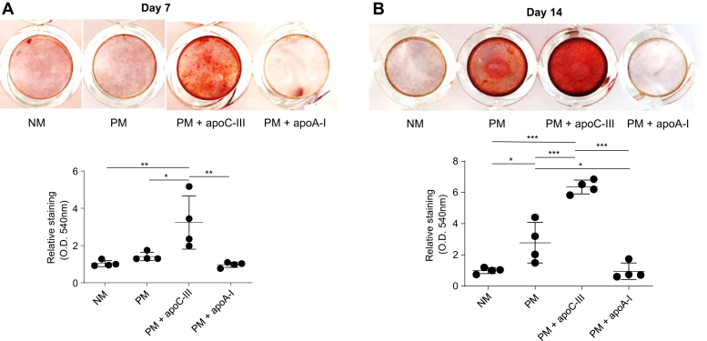
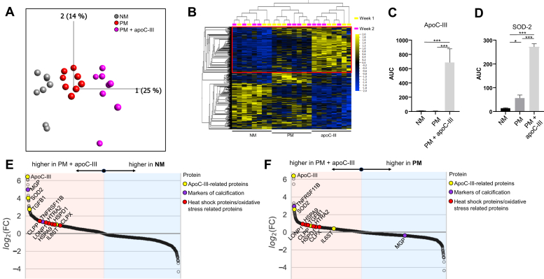
Calcific aortic valve disease (CAVD) occurs when subpopulations of valve cells undergo specific differentiation pathways, promoting tissue fibrosis and calcification. Lipoprotein particles carry oxidized lipids that promote valvular disease, but low-density lipoprotein-lowering therapies have failed in clinical trials, and there are currently no pharmacological interventions available for this disease. Apolipoproteins are known promoters of atherosclerosis, but whether they possess pathogenic properties in CAVD is less clear. To search for a possible link, we assessed 12 apolipoproteins in nonfibrotic/noncalcific and fibrotic/calcific aortic valve tissues by proteomics and immunohistochemistry to understand if they were enriched in calcified areas. Eight apolipoproteins (apoA-I, apoA-II, apoA-IV, apoB, apoC-III, apoD, apoL-I, and apoM) were enriched in the calcific versus nonfibrotic/noncalcific tissues. Apo(a), apoB, apoC-III, apoE, and apoJ localized within the disease-prone fibrosa and colocalized with calcific regions as detected by immunohistochemistry. Circulating apoC-III on lipoprotein(a) is a potential biomarker of aortic stenosis incidence and progression, but whether apoC-III also induces aortic valve calcification is unknown. We found that apoC-III was increased in fibrotic and calcific tissues and observed within the calcification-prone fibrosa layer as well as around calcification. In addition, we showed that apoC-III induced calcification in primary human valvular cell cultures via a mitochondrial dysfunction/inflammation-mediated pathway. This study provides a first assessment of a broad array of apolipoproteins in CAVD tissues, demonstrates that specific apolipoproteins associate with valvular calcification, and implicates apoC-III as an active and modifiable driver of CAVD beyond its potential role as a biomarker.
钙化性主动脉瓣疾病 (CAVD) 发生于瓣膜细胞的亚群经历特定的分化途径时,促进组织纤维化和钙化。脂蛋白颗粒携带促进瓣膜疾病的氧化脂质,但降低 LDL 的疗法在临床试验中失败了,目前没有针对这种疾病的药理学干预措施。载脂蛋白是动脉粥样硬化的已知促进剂,但它们在 CAVD 中是否具有致病性尚不清楚。为了寻找可能的联系,我们通过蛋白质组学和免疫组织化学评估了非纤维化/非钙化和纤维化/钙化主动脉瓣组织中的 12 种载脂蛋白,以了解它们是否在钙化区域富集。八种载脂蛋白(apoA-I、apoA-II、apoA-IV、apoB、apoC-III、apoD、apoL-I 和 apoM)在钙化与非纤维化/非钙化组织中富集。apo(a)、apoB、apoC-III、apoE 和 apoJ 在免疫组织化学检测到的易患病的纤维层中定位于病变部位,并与钙化区域共定位。脂蛋白 (a) 上的循环 apoC-III 是主动脉瓣狭窄发生率和进展的潜在生物标志物,但 apoC-III 是否也诱导主动脉瓣钙化尚不清楚。我们发现 apoC-III 在纤维化和钙化组织中增加,并在易钙化的纤维层内以及钙化周围观察到。此外,我们表明 apoC-III 通过线粒体功能障碍/炎症介导的途径诱导原代人瓣膜细胞培养物中的钙化。这项研究首次评估了 CAVD 组织中的广泛载脂蛋白,表明特定的载脂蛋白与瓣膜钙化相关,并暗示 apoC-III 是 CAVD 的一种活跃且可修饰的驱动因素,超出其作为生物标志物的潜在作用。