Sen Kuntal, Whitehead Matthew T, Gropman Andrea L
Division of Neurogenetics and Developmental Pediatrics, Center for Neuroscience and Behavioral Medicine, Children's National Hospital, Washington, DC, USA.
Department of Radiology, Children's National Hospital, Washington, DC, USA.
Transl Sci Rare Dis. 2020 Aug 3;5(1-2):87-95. doi: 10.3233/TRD-200048.
Urea cycle-related brain disease may take on variable neuroimaging manifestations, ranging from normal to abnormal with or without a signature appearance. In the past, we have described the usefulness of multimodal imaging in identifying biomarkers of neuronal injury in UCD patients. In this study, we report unique findings in an adolescent male with neonatal-onset OTC deficiency after an episode of hyperammonemia.
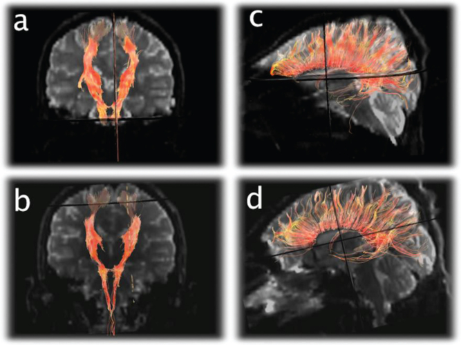
Multiplanar, multisequence MR imaging (T1WI, T2WI, T2 FLAIR, diffusion weighted images and gradient echo) of the brain was performed on seven separate occasions over the course following the acute illness; first five exams were performed within 28 days of admission and the final two exams were performed approximately 3 and 5 months later.
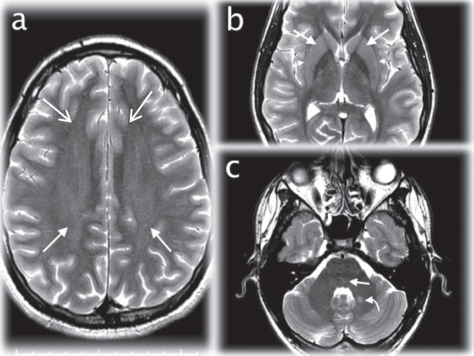
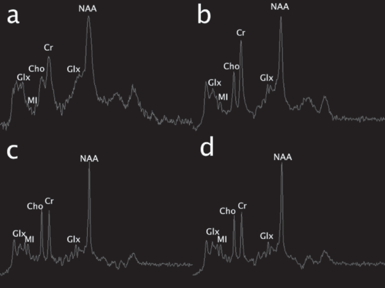
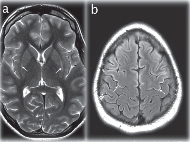
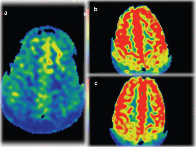
1.The initial MR revealed increased signal on T2WI in the basal ganglia, claustrum and frontoparietal white matter; which remained stable over time. By the 5th exam, signal changes had developed in frontal cortex; reflecting permanent injury. 2. DTI tractography of the corticospinal tracts displayed revealed diminution of the number of projectional and commissural fibers over time. 3. Blood flow measurements demonstrated hypoperfusion on the fifth exams followed by hyperperfusion on the final two studies. 4. MR spectroscopy demonstrated that glutamine was elevated during hyperammonemia with myoinositol reduction, reflecting osmotic buffering.
This particular multimodal magnetic resonance neuroimaging showed novel, temporally specific manifestations over the disease course in OTC deficiency. This prospective imaging study expands our understanding of the effect of hyperammonemia on the structure and biochemistry of the nervous system.
尿素循环相关脑疾病可能呈现出多种神经影像学表现,从正常到异常,有或无特征性表现。过去,我们已经描述了多模态成像在识别尿素循环障碍(UCD)患者神经元损伤生物标志物方面的作用。在本研究中,我们报告了一名青少年男性在新生儿期发病的鸟氨酸氨甲酰基转移酶(OTC)缺乏症患者发生高氨血症后的独特发现。
在急性疾病后的病程中,分七次对大脑进行多平面、多序列磁共振成像(T1加权成像、T2加权成像、液体衰减反转恢复序列、扩散加权成像和梯度回波)检查;前五次检查在入院后28天内进行,最后两次检查在大约3个月和5个月后进行。
这种特定的多模态磁共振神经成像在OTC缺乏症的病程中显示出新颖的、具有时间特异性的表现。这项前瞻性成像研究扩展了我们对高氨血症对神经系统结构和生物化学影响的理解。