Forghani Anoosha, Koduru Srinivas V, Chen Cong, Leberfinger Ashley N, Ravnic Dino J, Hayes Daniel J
Department of Biomedical Engineering, Millennium Science Complex, Pennsylvania State University, University Park, Pennsylvania, USA.
Department of Surgery, College of Medicine, Penn State Health Milton S. Hershey Medical Center, Hershey, Pennsylvania, USA.
Regen Eng Transl Med. 2020 Mar;6(1):101-110. doi: 10.1007/s40883-019-00093-7. Epub 2019 Mar 15.
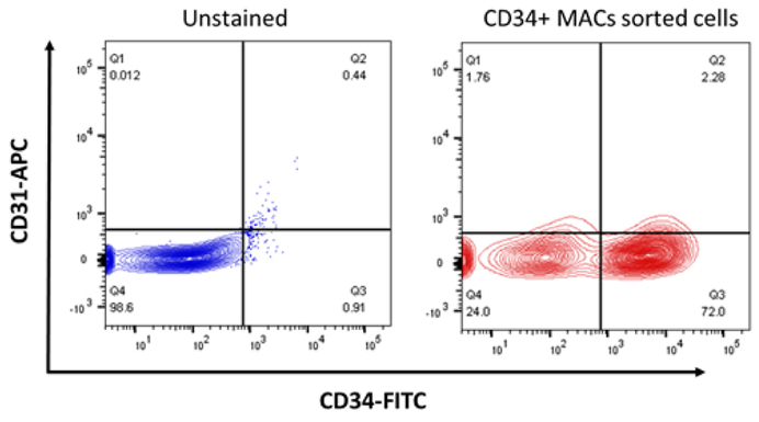
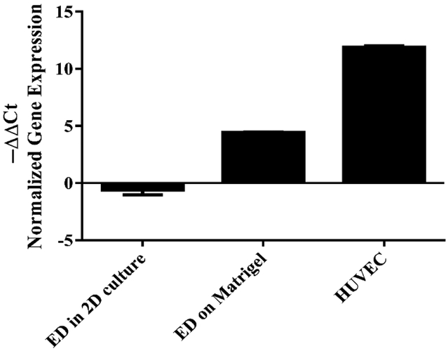
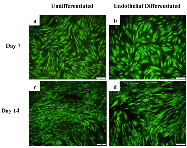
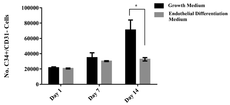
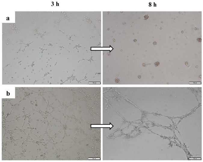
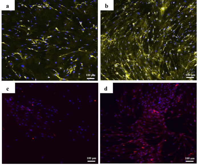
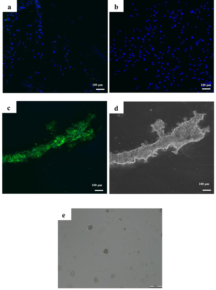
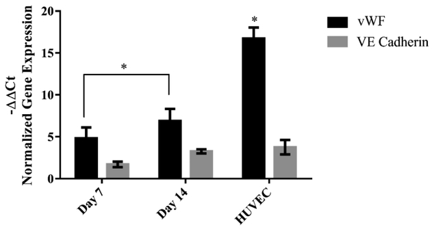
In this study, CD34/CD31 progenitor cells were isolated from the stromal vascular fraction (SVF) of adipose tissue using magnetic activated cell sorting. The endothelial differentiation capability of these cells was evaluated by culturing them in vascular endothelial growth factor (VEGF) induced medium for 14 days. Viability, proliferation, differentiation and tube formation of these cells were evaluated. Cell viability study revealed that both undifferentiated and endothelial differentiated cells remained healthy for 14 days. However, the proliferation rate was higher in undifferentiated cells compared to endothelial differentiated ones. Upregulation of endothelial characteristic genes (Von Willebrand Factor (vWF) and VE Cadherin) was observed in 2D culture. However, PECAM (CD31) was only found to be upregulated after the cells had formed tube-like structures in 3D Matrigel culture. These results indicate that adipose derived CD34/CD31 cells when cultured in VEGF induced medium, are capable differentiation into endothelial-like lineages. Tube formation of the cells started 3h after seeding the cells on Matrigel and formed more stable and connected network 24 h post seeding in presence of VEGF.
在本研究中,使用磁激活细胞分选技术从脂肪组织的基质血管成分(SVF)中分离出CD34/CD31祖细胞。通过在血管内皮生长因子(VEGF)诱导培养基中培养这些细胞14天来评估其内皮分化能力。对这些细胞的活力、增殖、分化和管形成进行了评估。细胞活力研究表明,未分化细胞和内皮分化细胞在14天内均保持健康。然而,未分化细胞的增殖率高于内皮分化细胞。在二维培养中观察到内皮特征基因(血管性血友病因子(vWF)和VE钙黏蛋白)的上调。然而,只有在细胞在三维基质胶培养中形成管状结构后,才发现血小板内皮细胞黏附分子(CD31)上调。这些结果表明,脂肪来源的CD34/CD31细胞在VEGF诱导培养基中培养时能够分化为内皮样谱系。将细胞接种到基质胶上3小时后开始形成管,在有VEGF的情况下接种24小时后形成更稳定且相互连接的网络。