Lu Edward S, Cui Ying, Le Rongrong, Zhu Ying, Wang Jay C, Laíns Inês, Katz Raviv, Lu Yifan, Zeng Rebecca, Garg Itika, Wu David M, Eliott Dean, Vavvas Demetrios G, Husain Deeba, Miller Joan W, Kim Leo A, Miller John B
Retina Service, Department of Ophthalmology, Massachusetts Eye and Ear, Harvard Medical School, Boston, Massachusetts, USA.
Harvard Retinal Imaging Lab, Massachusetts Eye and Ear, Boston, Massachusetts, USA.
Br J Ophthalmol. 2022 Apr;106(4):534-539. doi: 10.1136/bjophthalmol-2020-317983. Epub 2020 Dec 21.
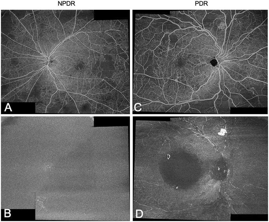
To compare the efficacy of diabetic retinal neovascularisation (NV) detection using the widefield swept-source optical coherence tomography angiography (WF SS-OCTA) vitreoretinal interface (VRI) Angio slab and SS-OCT VRI Structure slab.
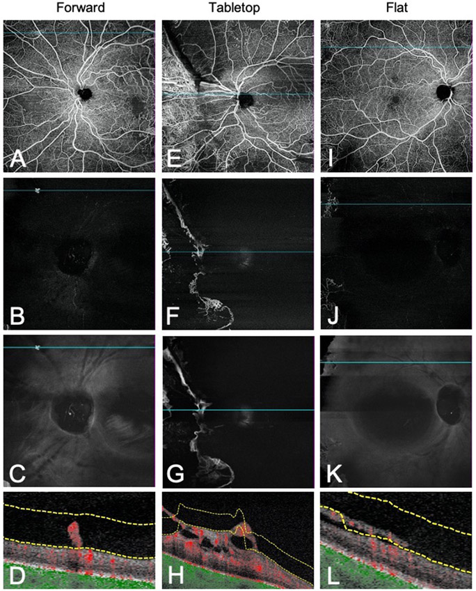
A prospective, observational study was performed at Massachusetts Eye and Ear from January 2019 to June 2020. Patients with proliferative diabetic retinopathy (PDR), patients with non-proliferative diabetic retinopathy and patients with diabetes but without diabetic retinopathy were included. All patients were imaged with WF SS-OCTA using the 12×12 mm Angio scan protocol centred on the fovea and optic disc. The en-face SS-OCTA VRI Angio slab and SS-OCT VRI Structure slab were evaluated for the presence or absence of NV. SS-OCTA B-scan was used to classify NV according to cross-sectional morphology (forward, tabletop or flat). All statistical analyses were performed using SPSS V.26.0.
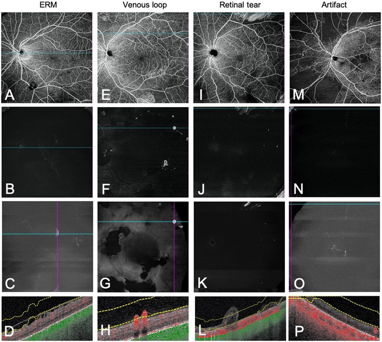
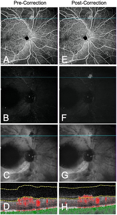
One hundred and forty-two eyes of 89 participants were included in the study. VRI Angio detected NV at higher rates compared with VRI Structure (p<0.05). Combining VRI Angio and Structure improved detection rates compared with VRI Angio alone (p<0.05). Due to segmentation errors of the internal limiting membrane, NV with flat morphological classification had lower rates of detection on VRI Angio compared with NV with forward and tabletop morphology (p<0.05).
WF SS-OCTA 12×12 mm VRI Angio and SS-OCT VRI Structure imaging centred on the fovea and optic disc detected NV with high sensitivity and low false positives. The VRI slab may be useful to diagnose and monitor PDR in clinical practice.
比较使用广角扫频源光学相干断层扫描血管造影(WF SS-OCTA)玻璃体视网膜界面(VRI)血管造影平板和SS-OCT VRI结构平板检测糖尿病视网膜新生血管(NV)的效果。
2019年1月至2020年6月在马萨诸塞州眼耳医院进行了一项前瞻性观察研究。纳入增殖性糖尿病视网膜病变(PDR)患者、非增殖性糖尿病视网膜病变患者以及患有糖尿病但无糖尿病视网膜病变的患者。所有患者均使用WF SS-OCTA以黄斑中心凹和视盘为中心的12×12毫米血管造影扫描协议进行成像。评估正面SS-OCTA VRI血管造影平板和SS-OCT VRI结构平板上NV的有无。使用SS-OCTA B扫描根据横截面形态(向前、桌面状或扁平状)对NV进行分类。所有统计分析均使用SPSS V.26.0进行。
89名参与者的142只眼纳入研究。与VRI结构相比,VRI血管造影检测NV的率更高(p<0.05)。与单独使用VRI血管造影相比,联合使用VRI血管造影和结构提高了检测率(p<0.05)。由于内界膜分割错误,与具有向前和桌面状形态的NV相比,具有扁平形态分类的NV在VRI血管造影上的检测率较低(p<0.05)。
以黄斑中心凹和视盘为中心的WF SS-OCTA 12×12毫米VRI血管造影和SS-OCT VRI结构成像检测NV具有高灵敏度和低假阳性率。VRI平板在临床实践中可能有助于诊断和监测PDR。