• 文献检索
  • 文档翻译
  • 深度研究
  • 学术资讯
  • Suppr Zotero 插件Zotero 插件
  • 邀请有礼
  • 套餐&价格
  • 历史记录
应用&插件
Suppr Zotero 插件Zotero 插件浏览器插件Mac 客户端Windows 客户端微信小程序
定价
高级版会员购买积分包购买API积分包
服务
文献检索文档翻译深度研究API 文档MCP 服务
关于我们
关于 Suppr公司介绍联系我们用户协议隐私条款
关注我们

Suppr 超能文献

核心技术专利:CN118964589B侵权必究
粤ICP备2023148730 号-1Suppr @ 2026

文献检索

告别复杂PubMed语法,用中文像聊天一样搜索,搜遍4000万医学文献。AI智能推荐,让科研检索更轻松。

立即免费搜索

文件翻译

保留排版,准确专业,支持PDF/Word/PPT等文件格式,支持 12+语言互译。

免费翻译文档

深度研究

AI帮你快速写综述,25分钟生成高质量综述,智能提取关键信息,辅助科研写作。

立即免费体验

角膜共焦显微镜检查与定量感觉测试及神经传导检查在诊断和分层糖尿病性周围神经病变严重程度方面的比较

Corneal confocal microscopy compared with quantitative sensory testing and nerve conduction for diagnosing and stratifying the severity of diabetic peripheral neuropathy.

作者信息

Ferdousi Maryam, Kalteniece Alise, Azmi Shazli, Petropoulos Ioannis N, Worthington Anne, D'Onofrio Luca, Dhage Shaishav, Ponirakis Georgios, Alam Uazman, Marshall Andrew, Faber Catharina G, Lauria Giuseppe, Soran Handrean, Malik Rayaz A

机构信息

Division of Cardiovascular Sciences, Cardiac Centre, Faculty of Biology, Medicine and Health, The University of Manchester, Manchester, UK.

Research Division, Weill Cornell Medical College in Qatar, Doha, Qatar.

出版信息

BMJ Open Diabetes Res Care. 2020 Dec;8(2). doi: 10.1136/bmjdrc-2020-001801.

DOI:10.1136/bmjdrc-2020-001801
PMID:33355206
原文链接:https://pmc.ncbi.nlm.nih.gov/articles/PMC7754626/
Abstract

INTRODUCTION

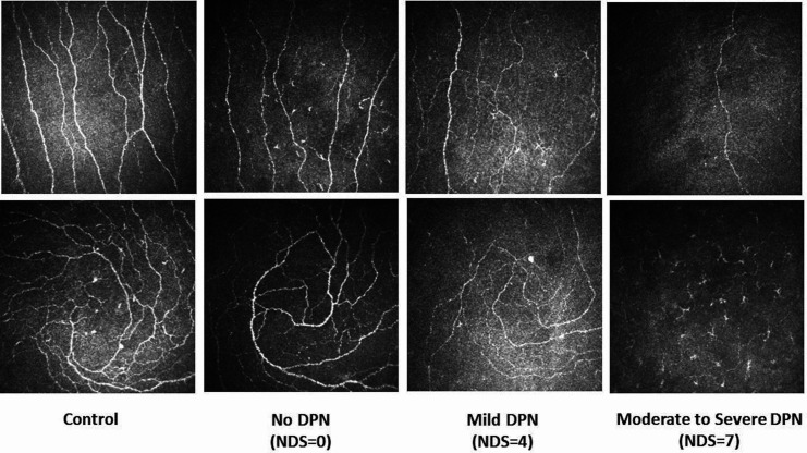
Diabetic neuropathy can be diagnosed and assessed using a number of techniques including corneal confocal microscopy (CCM).

RESEARCH DESIGN AND METHODS

We have undertaken quantitative sensory testing, nerve conduction studies and CCM in 143 patients with type 1 and type 2 diabetes without neuropathy (n=51), mild neuropathy (n=47) and moderate to severe neuropathy (n=45) and age-matched controls (n=30).

RESULTS

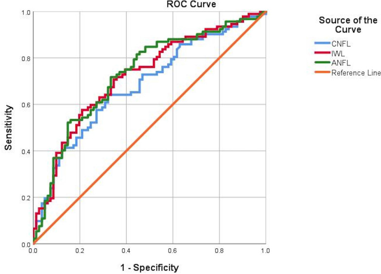
Vibration perception threshold (p<0.0001), warm perception threshold (WPT) (p<0.001), sural nerve conduction velocity (SNCV) (p<0.001), corneal nerve fiber density (CNFD) (p<0.0001), corneal nerve branch density (CNBD) (p<0.0001), corneal nerve fiber length (CNFL) (p=0.002), inferior whorl length (IWL) (p=0.0001) and average nerve fiber length (ANFL) (p=0.0001) showed a progressive abnormality with increasing severity of diabetic neuropathy. Receiver operating characteristic curve analysis for the diagnosis of diabetic neuropathy showed comparable performance in relation to the area under the curve (AUC) but differing sensitivities and specificities for vibration perception threshold (AUC 0.79, sensitivity 55%, specificity 90%), WPT (AUC 0.67, sensitivity 50%, specificity 76%), cold perception threshold (AUC 0.64, sensitivity 80%, specificity 47%), SNCV (AUC 0.70, sensitivity 76%, specificity 54%), CNFD (AUC 0.71, sensitivity 58%, specificity 83%), CNBD (AUC 0.70, sensitivity 69%, specificity 65%), CNFL (AUC 0.68, sensitivity 64%, specificity 67%), IWL (AUC 0.72, sensitivity 70%, specificity 65%) and ANFL (AUC 0.72, sensitivity 71%, specificity 66%).

CONCLUSION

This study shows that CCM identifies early and progressive corneal nerve loss at the inferior whorl and central cornea and has comparable utility with quantitative sensory testing and nerve conduction in the diagnosis of diabetic neuropathy.

摘要

引言

糖尿病性神经病变可通过多种技术进行诊断和评估,包括角膜共焦显微镜检查(CCM)。

研究设计与方法

我们对143例1型和2型糖尿病患者进行了定量感觉测试、神经传导研究和CCM检查,这些患者无神经病变(n = 51)、轻度神经病变(n = 47)、中度至重度神经病变(n = 45),并设有年龄匹配的对照组(n = 30)。

结果

振动觉阈值(p<0.0001)、温觉阈值(WPT)(p<0.001)、腓肠神经传导速度(SNCV)(p<0.001)、角膜神经纤维密度(CNFD)(p<0.0001)、角膜神经分支密度(CNBD)(p<0.0001)、角膜神经纤维长度(CNFL)(p = 0.002)、下象限长度(IWL)(p = 0.0001)和平均神经纤维长度(ANFL)(p = 0.0001)随着糖尿病性神经病变严重程度的增加呈现出逐渐加重的异常。糖尿病性神经病变诊断的受试者工作特征曲线分析显示,各指标在曲线下面积(AUC)方面表现相当,但在振动觉阈值(AUC 0.79,灵敏度55%,特异度90%)、WPT(AUC 0.67,灵敏度50%,特异度76%)、冷觉阈值(AUC 0.64,灵敏度80%,特异度47%)、SNCV(AUC 0.70,灵敏度76%,特异度54%)、CNFD(AUC 0.71,灵敏度58%,特异度83%)、CNBD(AUC 0.70,灵敏度69%,特异度65%)、CNFL(AUC 0.68,灵敏度64%,特异度67%)、IWL(AUC 0.72,灵敏度70%,特异度65%)和ANFL(AUC 0.72,灵敏度71%,特异度66%)方面的灵敏度和特异度有所不同。

结论

本研究表明,CCM可识别下象限和中央角膜早期及进行性角膜神经纤维丢失,在糖尿病性神经病变的诊断中与定量感觉测试和神经传导检查具有相当的效用。

https://cdn.ncbi.nlm.nih.gov/pmc/blobs/5cf5/7754626/e048e9956a75/bmjdrc-2020-001801f02.jpg
https://cdn.ncbi.nlm.nih.gov/pmc/blobs/5cf5/7754626/b5fca6c3075d/bmjdrc-2020-001801f01.jpg
https://cdn.ncbi.nlm.nih.gov/pmc/blobs/5cf5/7754626/e048e9956a75/bmjdrc-2020-001801f02.jpg
https://cdn.ncbi.nlm.nih.gov/pmc/blobs/5cf5/7754626/b5fca6c3075d/bmjdrc-2020-001801f01.jpg
https://cdn.ncbi.nlm.nih.gov/pmc/blobs/5cf5/7754626/e048e9956a75/bmjdrc-2020-001801f02.jpg

相似文献

1
Corneal confocal microscopy compared with quantitative sensory testing and nerve conduction for diagnosing and stratifying the severity of diabetic peripheral neuropathy.角膜共焦显微镜检查与定量感觉测试及神经传导检查在诊断和分层糖尿病性周围神经病变严重程度方面的比较
BMJ Open Diabetes Res Care. 2020 Dec;8(2). doi: 10.1136/bmjdrc-2020-001801.
2
The Inferior Whorl For Detecting Diabetic Peripheral Neuropathy Using Corneal Confocal Microscopy.用于通过角膜共焦显微镜检测糖尿病周围神经病变的下旋轮。
Invest Ophthalmol Vis Sci. 2015 Apr;56(4):2498-504. doi: 10.1167/iovs.14-15919.
3
Corneal confocal microscopy as a non-invasive test to assess diabetic peripheral neuropathy.角膜共焦显微镜作为一种非侵入性测试,用于评估糖尿病周围神经病变。
Diabetes Res Clin Pract. 2018 Feb;136:85-92. doi: 10.1016/j.diabres.2017.11.026. Epub 2017 Dec 6.
4
Diabetic Neuropathy Is Characterized by Progressive Corneal Nerve Fiber Loss in the Central and Inferior Whorl Regions.糖尿病性神经病的特征是中央和下象限区域的角膜神经纤维渐进性丧失。
Invest Ophthalmol Vis Sci. 2020 Mar 9;61(3):48. doi: 10.1167/iovs.61.3.48.
5
Greater corneal nerve loss at the inferior whorl is related to the presence of diabetic neuropathy and painful diabetic neuropathy.下象限角膜神经损失越大与糖尿病神经病变和痛性糖尿病神经病变的存在有关。
Sci Rep. 2018 Feb 19;8(1):3283. doi: 10.1038/s41598-018-21643-z.
6
Corneal confocal microscopy: A useful tool for diagnosis of small fiber neuropathy in type 2 diabetes.角膜共焦显微镜:诊断 2 型糖尿病小纤维神经病的有用工具。
J Diabetes Investig. 2021 Dec;12(12):2183-2189. doi: 10.1111/jdi.13616. Epub 2021 Jul 17.
7
Corneal sub-basal whorl-like nerve plexus: a landmark for early and follow-up evaluation in transthyretin familial amyloid polyneuropathy.角膜基底层涡状神经丛:转甲状腺素蛋白家族性淀粉样多发性神经病早期和随访评估的标志。
Eur J Neurol. 2021 Feb;28(2):630-638. doi: 10.1111/ene.14563. Epub 2020 Oct 24.
8
Corneal confocal microscopy identifies small fibre damage and progression of diabetic neuropathy.角膜共焦显微镜可识别小纤维损伤和糖尿病神经病变的进展。
Sci Rep. 2021 Jan 21;11(1):1859. doi: 10.1038/s41598-021-81302-8.
9
Small fiber neuropathy for assessment of disease severity in amyotrophic lateral sclerosis: corneal confocal microscopy findings.用于评估肌萎缩侧索硬化症疾病严重程度的小纤维神经病:角膜共聚焦显微镜检查结果。
Orphanet J Rare Dis. 2022 Jan 6;17(1):7. doi: 10.1186/s13023-021-02157-w.
10
Rapid automated diagnosis of diabetic peripheral neuropathy with in vivo corneal confocal microscopy.利用体内角膜共聚焦显微镜快速自动诊断糖尿病周围神经病变。
Invest Ophthalmol Vis Sci. 2014 Apr 3;55(4):2071-8. doi: 10.1167/iovs.13-13787.

引用本文的文献

1
Methods for the evaluation of corneal nerve fibres in diabetes mellitus by in vivo confocal microscopy: a scoping review.通过体内共聚焦显微镜评估糖尿病患者角膜神经纤维的方法:一项范围综述
BMJ Open. 2025 Sep 4;15(9):e098294. doi: 10.1136/bmjopen-2024-098294.
2
Ocular changes as potential biomarkers for early diagnosis of Alzheimer's disease.眼部变化作为阿尔茨海默病早期诊断的潜在生物标志物。
Alzheimers Dement. 2025 Aug;21(8):e70476. doi: 10.1002/alz.70476.
3
Deep-learning based analysis of in-vivo confocal microscopy images of the subbasal corneal nerve plexus' inferior whorl in patients with neuropathic corneal pain and dry eye disease.

本文引用的文献

1
Associations between acute glucose control and peripheral nerve structure and function in type 1 diabetes.1 型糖尿病患者急性血糖控制与周围神经结构和功能的关系。
Diabet Med. 2020 Sep;37(9):1553-1560. doi: 10.1111/dme.14306. Epub 2020 May 17.
2
Diabetic Neuropathy Is Characterized by Progressive Corneal Nerve Fiber Loss in the Central and Inferior Whorl Regions.糖尿病性神经病的特征是中央和下象限区域的角膜神经纤维渐进性丧失。
Invest Ophthalmol Vis Sci. 2020 Mar 9;61(3):48. doi: 10.1167/iovs.61.3.48.
3
Corneal confocal microscopy detects small nerve fibre damage in patients with painful diabetic neuropathy.
基于深度学习对神经性角膜疼痛和干眼症患者角膜基底下神经丛下旋的体内共聚焦显微镜图像进行分析。
Ocul Surf. 2024 Oct;34:241-246. doi: 10.1016/j.jtos.2024.08.002. Epub 2024 Aug 2.
4
Comparison of Novel Wide-Field In Vivo Corneal Confocal Microscopy With Skin Biopsy for Assessing Peripheral Neuropathy in Type 2 Diabetes.新型广角活体角膜共焦显微镜与皮肤活检评估 2 型糖尿病周围神经病变的比较。
Diabetes. 2023 Jul 1;72(7):908-917. doi: 10.2337/db22-0863.
5
Assessing Corneal Confocal Microscopy and Other Small Fiber Measures in Diabetic Polyneuropathy.评估糖尿病性多发性神经病中的角膜共焦显微镜和其他小纤维测量方法。
Neurology. 2023 Apr 18;100(16):e1680-e1690. doi: 10.1212/WNL.0000000000206902. Epub 2023 Feb 7.
6
Recent updates in the treatment of diabetic polyneuropathy.糖尿病性多发性神经病治疗的最新进展
Fac Rev. 2022 Oct 18;11:30. doi: 10.12703/r/11-30. eCollection 2022.
7
Continuous glucose monitoring reveals a novel association between duration and severity of hypoglycemia, and small nerve fiber injury in patients with diabetes.持续血糖监测揭示了糖尿病患者低血糖持续时间和严重程度与小神经纤维损伤之间的新关联。
Endocr Connect. 2022 Nov 14;11(12). doi: 10.1530/EC-22-0352. Print 2022 Dec 1.
8
Diabetic corneal neuropathy as a surrogate marker for diabetic peripheral neuropathy.糖尿病性角膜神经病变作为糖尿病周围神经病变的替代标志物。
Neural Regen Res. 2022 Oct;17(10):2172-2178. doi: 10.4103/1673-5374.327364.
9
Diabetes Distal Peripheral Neuropathy: Subtypes and Diagnostic and Screening Technologies.糖尿病远端周围神经病变:亚型和诊断及筛查技术。
J Diabetes Sci Technol. 2022 Mar;16(2):295-320. doi: 10.1177/19322968211035375. Epub 2022 Jan 7.
10
Therapeutic effect of opioid analgesics combined with non-steroidal anti-inflammatory drugs on peripheral neuropathy and its influence on inflammatory factors.阿片类镇痛药联合非甾体抗炎药对周围神经病变的治疗作用及其对炎症因子的影响
Am J Transl Res. 2021 Oct 15;13(10):11752-11757. eCollection 2021.
角膜共焦显微镜检测痛性糖尿病周围神经病患者的小纤维神经损伤。
Sci Rep. 2020 Feb 25;10(1):3371. doi: 10.1038/s41598-020-60422-7.
4
Corneal confocal microscopy: ready for prime time.角膜共焦显微镜:准备好进入黄金时期。
Clin Exp Optom. 2020 May;103(3):265-277. doi: 10.1111/cxo.12887. Epub 2019 Mar 4.
5
Corneal confocal microscopy for identification of diabetic sensorimotor polyneuropathy: a pooled multinational consortium study.角膜共焦显微镜在识别糖尿病感觉运动多发性神经病中的应用:一项多国家联合研究。
Diabetologia. 2018 Aug;61(8):1856-1861. doi: 10.1007/s00125-018-4653-8. Epub 2018 Jun 4.
6
Greater corneal nerve loss at the inferior whorl is related to the presence of diabetic neuropathy and painful diabetic neuropathy.下象限角膜神经损失越大与糖尿病神经病变和痛性糖尿病神经病变的存在有关。
Sci Rep. 2018 Feb 19;8(1):3283. doi: 10.1038/s41598-018-21643-z.
7
Corneal confocal microscopy is a rapid reproducible ophthalmic technique for quantifying corneal nerve abnormalities.角膜共焦显微镜检查是一种用于量化角膜神经异常的快速可重复的眼科技术。
PLoS One. 2017 Aug 17;12(8):e0183040. doi: 10.1371/journal.pone.0183040. eCollection 2017.
8
Diagnostic utility of corneal confocal microscopy and intra-epidermal nerve fibre density in diabetic neuropathy.角膜共焦显微镜检查和表皮内神经纤维密度在糖尿病神经病变中的诊断效用
PLoS One. 2017 Jul 18;12(7):e0180175. doi: 10.1371/journal.pone.0180175. eCollection 2017.
9
Diabetic Neuropathy: A Position Statement by the American Diabetes Association.糖尿病神经病变:美国糖尿病协会的立场声明
Diabetes Care. 2017 Jan;40(1):136-154. doi: 10.2337/dc16-2042.
10
The Reliability and Reproducibility of Corneal Confocal Microscopy in Children.儿童角膜共焦显微镜检查的可靠性和可重复性
Invest Ophthalmol Vis Sci. 2015 Aug;56(9):5636-40. doi: 10.1167/iovs.15-16995.